The coenzyme analogue adenosine 5-diphosphoribose displaces FAD in the active site of p-hydroxybenzoate hydroxylase. An x-ray crystallographic investigation.
van der Laan, J.M., Schreuder, H.A., Swarte, M.B., Wierenga, R.K., Kalk, K.H., Hol, W.G., Drenth, J.(1989) Biochemistry 28: 7199-7205
- PubMed: 2819062 Search on PubMed
- DOI: https://doi.org/10.1021/bi00444a011
- Primary Citation Related Structures:
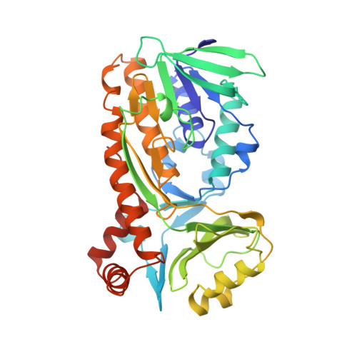
2PHH - PubMed Abstract:
p-Hydroxybenzoate hydroxylase (PHBH) is an NADPH-dependent enzyme. To locate the NADPH binding site, the enzyme was crystallized under anaerobic conditions in the presence of the substrate p-hydroxybenzoate, the coenzyme analogue adenosine 5-diphosphoribose (ADPR), and sodium dithionite. This yielded colorless crystals that were suitable for X-ray analysis. Diffraction data were collected up to 2.7-A resolution. A difference Fourier between data from these colorless crystals and data from yellow crystals of the enzyme-substrate complex showed that in the colorless crystals the flavin ring was absent. The adenosine 5'-diphosphate moiety, which is the common part between FAD and ADPR, was still present. After restrained least-squares refinement of the enzyme-substrate complex with the riboflavin omitted from the model, additional electron density appeared near the pyrophosphate, which indicated the presence of an ADPR molecule in the FAD binding site of PHBH. The complete ADPR molecule was fitted to the electron density, and subsequent least-squares refinement resulted in a final R factor of 16.8%. Replacement of bound FAD by ADPR was confirmed by equilibrium dialysis, where it was shown that ADPR can effectively remove FAD from the enzyme under mild conditions in 0.1 M potassium phosphate buffer, pH 8.0. The empty pocket left by the flavin ring is filled by solvent, leaving the architecture of the active site and the binding of the substrate largely unaffected.
- Laboratory of Chemical Physics, University of Groningen, The Netherlands.
Organizational Affiliation: