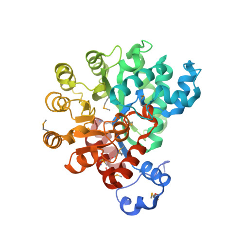
2PGR

Crystal structure of adenosine deaminase from Plasmodium vivax in complex with pentostatin


Experimental Data Snapshot

  • Method: X-RAY DIFFRACTION
  • Resolution: 2.30 Å
  • R-Value Free: 0.220 
  • R-Value Work: 0.168 
  • R-Value Observed: 0.171 

Starting Model: experimental
View more details

wwPDB Validation   3D Report Full Report


Ligand Structure Quality Assessment 


This is version 1.6 of the entry. See complete history


Literature

Structures of substrate- and inhibitor-bound adenosine deaminase from a human malaria parasite show a dramatic conformational change and shed light on drug selectivity.

Larson, E.T.Deng, W.Krumm, B.E.Napuli, A.Mueller, N.Van Voorhis, W.C.Buckner, F.S.Fan, E.Lauricella, A.DeTitta, G.Luft, J.Zucker, F.Hol, W.G.Verlinde, C.L.Merritt, E.A.

(2008) J Mol Biol 381: 975-988

  • DOI: https://doi.org/10.1016/j.jmb.2008.06.048
  • Primary Citation of Related Structures:  
    2PGF, 2PGR, 2QVN

  • PubMed Abstract: 

    Plasmodium and other apicomplexan parasites are deficient in purine biosynthesis, relying instead on the salvage of purines from their host environment. Therefore, interference with the purine salvage pathway is an attractive therapeutic target. The plasmodial enzyme adenosine deaminase (ADA) plays a central role in purine salvage and, unlike mammalian ADA homologs, has a further secondary role in methylthiopurine recycling. For this reason, plasmodial ADA accepts a wider range of substrates, as it is responsible for deamination of both adenosine and 5'-methylthioadenosine. The latter substrate is not accepted by mammalian ADA homologs. The structural basis for this natural difference in specificity between plasmodial and mammalian ADA has not been well understood. We now report crystal structures of Plasmodium vivax ADA in complex with adenosine, guanosine, and the picomolar inhibitor 2'-deoxycoformycin. These structures highlight a drastic conformational change in plasmodial ADA upon substrate binding that has not been observed for mammalian ADA enzymes. Further, these complexes illuminate the structural basis for the differential substrate specificity and potential drug selectivity between mammalian and parasite enzymes.


  • Organizational Affiliation

    Medical Structural Genomics of Pathogenic Protozoa Consortium, University of Washington, Seattle, WA 98195-7742, USA.


Macromolecules
Find similar proteins by:  (by identity cutoff)  |  3D Structure
Entity ID: 1
MoleculeChains Sequence LengthOrganismDetailsImage
adenosine deaminase371Plasmodium vivaxMutation(s): 0 
Gene Names: Pv111245
EC: 3.5.4.4 (PDB Primary Data), 3.5.4.31 (UniProt)
UniProt
Find proteins for A5KE01 (Plasmodium vivax (strain Salvador I))
Explore A5KE01 
Go to UniProtKB:  A5KE01
Entity Groups  
Sequence Clusters30% Identity50% Identity70% Identity90% Identity95% Identity100% Identity
UniProt GroupA5KE01
Sequence Annotations
Expand
  • Reference Sequence
Experimental Data & Validation

Experimental Data

  • Method: X-RAY DIFFRACTION
  • Resolution: 2.30 Å
  • R-Value Free: 0.220 
  • R-Value Work: 0.168 
  • R-Value Observed: 0.171 
  • Space Group: C 2 2 21
Unit Cell:
Length ( Å )Angle ( ˚ )
a = 143.488α = 90
b = 146.386β = 90
c = 50.034γ = 90
Software Package:
Software NamePurpose
DENZOdata reduction
SCALEPACKdata scaling
REFMACrefinement
PDB_EXTRACTdata extraction
Blu-Icedata collection
HKL-2000data reduction
HKL-2000data scaling
MOLREPphasing

Structure Validation

View Full Validation Report


Ligand Structure Quality Assessment 


Entry History 

Deposition Data

Revision History  (Full details and data files)

  • Version 1.0: 2007-04-24
    Type: Initial release
  • Version 1.1: 2007-09-24
    Changes: Version format compliance
  • Version 1.2: 2011-07-13
    Changes: Advisory, Version format compliance
  • Version 1.3: 2017-10-18
    Changes: Refinement description
  • Version 1.4: 2023-08-30
    Changes: Data collection, Database references, Derived calculations, Refinement description
  • Version 1.5: 2023-11-15
    Changes: Data collection
  • Version 1.6: 2024-10-30
    Changes: Structure summary