2PFG | pdb_00002pfg

Crystal structure of human CBR1 in complex with BiGF2.


Experimental Data Snapshot

  • Method: X-RAY DIFFRACTION
  • Resolution: 1.54 Å
  • R-Value Free: 
    0.164 (Depositor), 0.161 (DCC) 
  • R-Value Work: 
    0.112 (Depositor), 0.108 (DCC) 
  • R-Value Observed: 
    0.115 (Depositor) 

Starting Model: experimental
View more details

wwPDB Validation 3D Report Full Report

Validation slider image for 2PFG

Ligand Structure Quality Assessment 


This is version 2.0 of the entry. See complete history

Literature

Glutathione traps formaldehyde by formation of a bicyclo[4.4.1]undecane adduct.

Bateman, R.Rauh, D.Shokat, K.M.

(2007) Org Biomol Chem 5: 3363-3367

  • DOI: https://doi.org/10.1039/b707602a
  • Primary Citation Related Structures: 
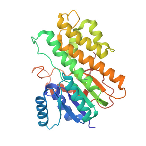
    2PFG

  • PubMed Abstract: 

    Glutathione forms complex reaction products with formaldehyde, which can be further modified through enzymatic modification. We studied the non-enzymatic reaction between glutathione and formaldehyde and identified a bicyclic complex containing two equivalents of formaldehyde and one glutathione molecule by protein X-ray crystallography (PDB accession number 2PFG). We have also used (1)H, (13)C and 2D NMR spectroscopy to confirm the structure of this unusual adduct. The key feature of this adduct is the involvement of the gamma-glutamyl alpha-amine and the Cys thiol in the formation of the bicyclic ring structure. These findings suggest that the structure of GSH allows for bi-dentate masking of the reactivity of formaldehyde. As this species predominates at near physiological pH values, we suggest this adduct may have biological significance.


  • Organizational Affiliation
    • Howard Hughes Medical Institute, Department of Cellular and Molecular Pharmacology, UCSF, 600 16th St., San Francisco, CA 94143-2280, USA.

Macromolecule Content 

  • Total Structure Weight: 31.52 kDa 
  • Atom Count: 2,690 
  • Modeled Residue Count: 274 
  • Deposited Residue Count: 276 
  • Unique protein chains: 1

Macromolecules

Find similar proteins by:|  3D Structure
Entity ID: 1
MoleculeChains  Sequence LengthOrganismDetailsImage
Carbonyl reductase [NADPH] 1276Homo sapiensMutation(s): 0 
Gene Names: CBR1CBRCRN
EC: 1.1.1.184 (PDB Primary Data), 1.1.1.189 (PDB Primary Data), 1.1.1.197 (PDB Primary Data), 1.1.1.196 (UniProt), 1.1.1.71 (UniProt)
UniProt & NIH Common Fund Data Resources
Find proteins for P16152 (Homo sapiens)
Explore P16152 
Go to UniProtKB:  P16152
PHAROS:  P16152
GTEx:  ENSG00000159228 
Entity Groups
Sequence Clusters30% Identity50% Identity70% Identity90% Identity95% Identity100% Identity
UniProt GroupP16152
Sequence Annotations
Expand
Reference Sequence

Small Molecules

Ligands 4 Unique
IDChains Name / Formula / InChI Key2D Diagram3D Interactions
NAP

Query on NAP


Download:Ideal Coordinates CCD File
C [auth A]NADP NICOTINAMIDE-ADENINE-DINUCLEOTIDE PHOSPHATE
C21 H28 N7 O17 P3
XJLXINKUBYWONI-NNYOXOHSSA-N
DDD

Query on DDD


Download:Ideal Coordinates CCD File
D [auth A](5R,10S)-5-{[(CARBOXYMETHYL)AMINO]CARBONYL}-7-OXO-3-THIA-1,6-DIAZABICYCLO[4.4.1]UNDECANE-10-CARBOXYLIC ACID
C12 H17 N3 O6 S
FNEMNSFAPZMNDP-YUMQZZPRSA-N
GOL

Query on GOL


Download:Ideal Coordinates CCD File
E [auth A]GLYCEROL
C3 H8 O3
PEDCQBHIVMGVHV-UHFFFAOYSA-N
CL

Query on CL


Download:Ideal Coordinates CCD File
B [auth A]CHLORIDE ION
Cl
VEXZGXHMUGYJMC-UHFFFAOYSA-M
Modified Residues  1 Unique
IDChains TypeFormula2D DiagramParent
M0H
Query on M0H
A
L-PEPTIDE LINKINGC4 H9 N O3 SCYS

Experimental Data & Validation

Experimental Data

  • Method: X-RAY DIFFRACTION
  • Resolution: 1.54 Å
  • R-Value Free:  0.164 (Depositor), 0.161 (DCC) 
  • R-Value Work:  0.112 (Depositor), 0.108 (DCC) 
  • R-Value Observed: 0.115 (Depositor) 
Space Group: P 21 21 21
Unit Cell:
Length ( Å )Angle ( ˚ )
a = 54.636α = 90
b = 59.884β = 90
c = 87.954γ = 90
Software Package:
Software NamePurpose
REFMACrefinement
HKL-2000data collection
HKL-2000data reduction
HKL-2000data scaling
AMoREphasing

Structure Validation

View Full Validation Report


Ligand Structure Quality Assessment 


Entry History 

Deposition Data

Revision History  (Full details and data files)

  • Version 1.0: 2007-09-25
    Type: Initial release
  • Version 1.1: 2011-07-13
    Changes: Non-polymer description, Version format compliance
  • Version 1.2: 2023-08-30
    Changes: Data collection, Database references, Derived calculations, Refinement description
  • Version 2.0: 2023-11-15
    Changes: Atomic model, Data collection, Derived calculations