2PFC | pdb_00002pfc

Structure of Mycobacterium tuberculosis Rv0098


Experimental Data Snapshot

  • Method: X-RAY DIFFRACTION
  • Resolution: 2.30 Å
  • R-Value Free: 
    0.273 (Depositor) 
  • R-Value Work: 
    0.235 (Depositor) 
  • R-Value Observed: 
    0.237 (Depositor) 

wwPDB Validation 3D Report Full Report

Validation slider image for 2PFC

This is version 1.2 of the entry. See complete history

Literature

Identification of a type III thioesterase reveals the function of an operon crucial for Mtb virulence.

Wang, F.Langley, R.Gulten, G.Wang, L.Sacchettini, J.C.

(2007) Chem Biol 14: 543-551

  • DOI: https://doi.org/10.1016/j.chembiol.2007.04.005
  • Primary Citation Related Structures: 
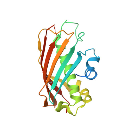
    2PFC

  • PubMed Abstract: 

    Rv0098 is part of an operon, Rv0096-Rv0101, from Mycobacterium tuberculosis (Mtb) that is essential for Mtb's survival in mouse macrophages. This operon also contains an acyl carrier protein and one of the only two nonribosomal peptide synthases in Mtb. Rv0098 is annotated in the genome as a hypothetical protein and was proposed to be an acyl-coenzyme A (CoA) dehydratase. The structure of Rv0098, together with subsequent biochemical analysis, indicated that Rv0098 is a long-chain fatty acyl-CoA thioesterase (FcoT). However, FcoT lacks a general base or a nucleophile that is always found in the catalytic site of type II and type I thioesterases, respectively. The active site of Mtb FcoT reveals the structural basis for its substrate specificity for long-chain acyl-CoA and allows us to propose a catalytic mechanism for the enzyme. The characterization of Mtb FcoT provides a putative function of this operon that is crucial for Mtb pathogenicity.


  • Organizational Affiliation
    • Department of Biochemistry and Biophysics, Texas A&M University, College Station, TX 77843, USA.

Macromolecule Content 

  • Total Structure Weight: 20.81 kDa 
  • Atom Count: 1,310 
  • Modeled Residue Count: 160 
  • Deposited Residue Count: 183 
  • Unique protein chains: 1

Macromolecules

Find similar proteins by:|  3D Structure
Entity ID: 1
MoleculeChains  Sequence LengthOrganismDetailsImage
Hypothetical protein Rv0098/MT0107183Mycobacterium tuberculosisMutation(s): 0 
EC: 3.1.2.2 (UniProt), 4.3.2.11 (UniProt), 3.1.2 (UniProt)
UniProt
Find proteins for P9WM67 (Mycobacterium tuberculosis (strain ATCC 25618 / H37Rv))
Explore P9WM67 
Go to UniProtKB:  P9WM67
Entity Groups
Sequence Clusters30% Identity50% Identity70% Identity90% Identity95% Identity100% Identity
UniProt GroupP9WM67
Sequence Annotations
Expand
Reference Sequence

Small Molecules

Ligands 1 Unique
IDChains Name / Formula / InChI Key2D Diagram3D Interactions
PLM

Query on PLM


Download:Ideal Coordinates CCD File
B [auth A]PALMITIC ACID
C16 H32 O2
IPCSVZSSVZVIGE-UHFFFAOYSA-N

Experimental Data & Validation

Experimental Data

  • Method: X-RAY DIFFRACTION
  • Resolution: 2.30 Å
  • R-Value Free:  0.273 (Depositor) 
  • R-Value Work:  0.235 (Depositor) 
  • R-Value Observed: 0.237 (Depositor) 
Space Group: I 21 3
Unit Cell:
Length ( Å )Angle ( ˚ )
a = 101.767α = 90
b = 101.767β = 90
c = 101.767γ = 90
Software Package:
Software NamePurpose
REFMACrefinement
HKL-2000data reduction
HKL-2000data scaling
SHARPphasing

Structure Validation

View Full Validation Report


Entry History 

Deposition Data

Revision History  (Full details and data files)

  • Version 1.0: 2008-04-08
    Type: Initial release
  • Version 1.1: 2011-07-13
    Changes: Version format compliance
  • Version 1.2: 2024-10-16
    Changes: Data collection, Database references, Derived calculations, Structure summary