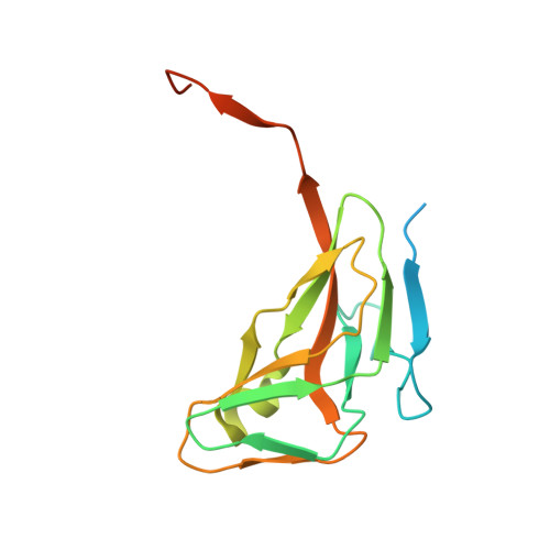
2PC5

Native crystal structure analysis on Arabidopsis dUTPase


Experimental Data Snapshot

  • Method: X-RAY DIFFRACTION
  • Resolution: 2.20 Å
  • R-Value Free: 0.251 
  • R-Value Work: 0.182 
  • R-Value Observed: 0.185 

Starting Model: experimental
View more details

wwPDB Validation   3D Report Full Report


This is version 1.2 of the entry. See complete history


Literature

Purification, crystallization and preliminary crystallographic analysis of deoxyuridine triphosphate nucleotidohydrolase from Arabidopsis thaliana.

Bajaj, M.Moriyama, H.

(2007) Acta Crystallogr Sect F Struct Biol Cryst Commun 63: 409-411

  • DOI: https://doi.org/10.1107/S1744309107016004
  • Primary Citation of Related Structures:  
    2PC5

  • PubMed Abstract: 

    The deoxyuridine triphosphate nucleotidohydrolase gene from Arabidopsis thaliana was expressed and the gene product was purified. Crystallization was performed by the hanging-drop vapour-diffusion method at 298 K using 2 M ammonium sulfate as the precipitant. X-ray diffraction data were collected to 2.2 A resolution using Cu K alpha radiation. The crystal belongs to the orthorhombic space group P2(1)2(1)2(1), with unit-cell parameters a = 69.90, b = 70.86 A, c = 75.55 A. Assuming the presence of a trimer in the asymmetric unit, the solvent content was 30%, with a V(M) of 1.8 A3 Da(-1).


  • Organizational Affiliation

    School of Biological Sciences, University of Nebraska-Lincoln, Manter Hall, Lincoln, Nebraska 68588-0304, USA.


Macromolecules
Find similar proteins by:  (by identity cutoff)  |  3D Structure
Entity ID: 1
MoleculeChains Sequence LengthOrganismDetailsImage
DUTP pyrophosphatase-like protein
A, B, C
169Arabidopsis thalianaMutation(s): 0 
Gene Names: T6H20.30
EC: 3.6.1.23
UniProt
Find proteins for Q9STG6 (Arabidopsis thaliana)
Explore Q9STG6 
Go to UniProtKB:  Q9STG6
Entity Groups  
Sequence Clusters30% Identity50% Identity70% Identity90% Identity95% Identity100% Identity
UniProt GroupQ9STG6
Sequence Annotations
Expand
  • Reference Sequence
Small Molecules
Ligands 1 Unique
IDChains Name / Formula / InChI Key2D Diagram3D Interactions
MG
Query on MG

Download Ideal Coordinates CCD File 
D [auth A]MAGNESIUM ION
Mg
JLVVSXFLKOJNIY-UHFFFAOYSA-N
Experimental Data & Validation

Experimental Data

  • Method: X-RAY DIFFRACTION
  • Resolution: 2.20 Å
  • R-Value Free: 0.251 
  • R-Value Work: 0.182 
  • R-Value Observed: 0.185 
  • Space Group: P 21 21 21
Unit Cell:
Length ( Å )Angle ( ˚ )
a = 69.903α = 90
b = 70.86β = 90
c = 75.555γ = 90
Software Package:
Software NamePurpose
REFMACrefinement
CrystalCleardata collection
HKL-2000data reduction
HKL-2000data scaling
MOLREPphasing

Structure Validation

View Full Validation Report


Entry History 

Deposition Data

Revision History  (Full details and data files)

  • Version 1.0: 2008-02-12
    Type: Initial release
  • Version 1.1: 2011-07-13
    Changes: Derived calculations, Version format compliance
  • Version 1.2: 2023-08-30
    Changes: Data collection, Database references, Derived calculations, Refinement description