2PAN | pdb_00002pan

Crystal structure of E. coli glyoxylate carboligase


Experimental Data Snapshot

  • Method: X-RAY DIFFRACTION
  • Resolution: 2.70 Å
  • R-Value Free: 
    0.253 (Depositor) 
  • R-Value Work: 
    0.219 (Depositor) 
  • R-Value Observed: 
    0.220 (Depositor) 

wwPDB Validation 3D Report Full Report

Validation slider image for 2PAN

Ligand Structure Quality Assessment 


This is version 2.1 of the entry. See complete history

Literature

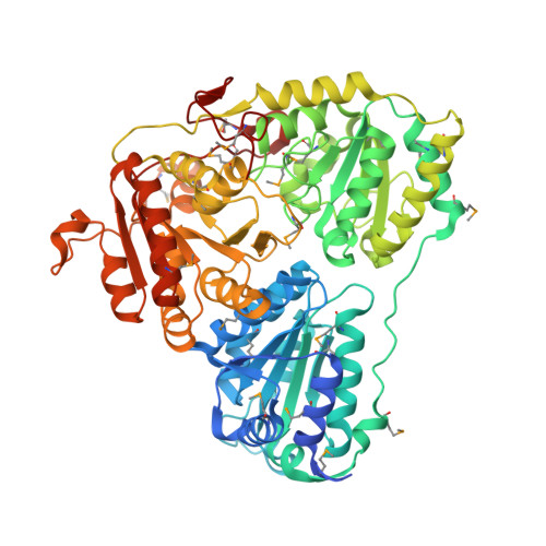
Glyoxylate carboligase lacks the canonical active site glutamate of thiamine-dependent enzymes.

Kaplun, A.Binshtein, E.Vyazmensky, M.Steinmetz, A.Barak, Z.Chipman, D.M.Tittmann, K.Shaanan, B.

(2008) Nat Chem Biol 4: 113-118

  • DOI: https://doi.org/10.1038/nchembio.62
  • Primary Citation Related Structures: 
    2PAN

  • PubMed Abstract: 

    Thiamine diphosphate (ThDP), a derivative of vitamin B1, is an enzymatic cofactor whose special chemical properties allow it to play critical mechanistic roles in a number of essential metabolic enzymes. It has been assumed that all ThDP-dependent enzymes exploit a polar interaction between a strictly conserved glutamate and the N1' of the ThDP moiety. The crystal structure of glyoxylate carboligase challenges this paradigm by revealing that valine replaces the conserved glutamate. Through kinetic, spectroscopic and site-directed mutagenesis studies, we show that although this extreme change lowers the rate of the initial step of the enzymatic reaction, it ensures efficient progress through subsequent steps. Glyoxylate carboligase thus provides a unique illustration of the fine tuning between catalytic stages imposed during evolution on enzymes catalyzing multistep processes.


  • Organizational Affiliation
    • Department of Life Sciences, Ben-Gurion University of the Negev, 1 Ben-Gurion Avenue, Beer-Sheva 84105, Israel.

Macromolecule Content 

  • Total Structure Weight: 419.29 kDa 
  • Atom Count: 28,107 
  • Modeled Residue Count: 3,552 
  • Deposited Residue Count: 3,696 
  • Unique protein chains: 1

Macromolecules

Find similar proteins by:|  3D Structure
Entity ID: 1
MoleculeChains  Sequence LengthOrganismDetailsImage
Glyoxylate carboligase
A, B, C, D, E
A, B, C, D, E, F
616Escherichia coliMutation(s): 0 
Gene Names: gcl
EC: 4.1.1.47
UniProt
Find proteins for P0AEP7 (Escherichia coli (strain K12))
Explore P0AEP7 
Go to UniProtKB:  P0AEP7
Entity Groups
Sequence Clusters30% Identity50% Identity70% Identity90% Identity95% Identity100% Identity
UniProt GroupP0AEP7
Sequence Annotations
Expand
Reference Sequence

Small Molecules

Ligands 5 Unique
IDChains Name / Formula / InChI Key2D Diagram3D Interactions
FAD

Query on FAD


Download:Ideal Coordinates CCD File
BA [auth E]
GA [auth F]
I [auth A]
O [auth B]
S [auth C]
BA [auth E],
GA [auth F],
I [auth A],
O [auth B],
S [auth C],
W [auth D]
FLAVIN-ADENINE DINUCLEOTIDE
C27 H33 N9 O15 P2
VWWQXMAJTJZDQX-UYBVJOGSSA-N
TPP

Query on TPP


Download:Ideal Coordinates CCD File
CA [auth E]
HA [auth F]
J [auth A]
P [auth B]
T [auth C]
CA [auth E],
HA [auth F],
J [auth A],
P [auth B],
T [auth C],
X [auth D]
THIAMINE DIPHOSPHATE
C12 H19 N4 O7 P2 S
AYEKOFBPNLCAJY-UHFFFAOYSA-O
1PE

Query on 1PE


Download:Ideal Coordinates CCD File
L [auth A]PENTAETHYLENE GLYCOL
C10 H22 O6
JLFNLZLINWHATN-UHFFFAOYSA-N
DTT

Query on DTT


Download:Ideal Coordinates CCD File
DA [auth E]
IA [auth F]
K [auth A]
Q [auth B]
U [auth C]
DA [auth E],
IA [auth F],
K [auth A],
Q [auth B],
U [auth C],
Y [auth D]
2,3-DIHYDROXY-1,4-DITHIOBUTANE
C4 H10 O2 S2
VHJLVAABSRFDPM-IMJSIDKUSA-N
MG

Query on MG


Download:Ideal Coordinates CCD File
AA [auth E]
EA [auth F]
FA [auth F]
G [auth A]
H [auth A]
AA [auth E],
EA [auth F],
FA [auth F],
G [auth A],
H [auth A],
M [auth B],
N [auth B],
R [auth C],
V [auth D],
Z [auth E]
MAGNESIUM ION
Mg
JLVVSXFLKOJNIY-UHFFFAOYSA-N
Modified Residues  1 Unique
IDChains TypeFormula2D DiagramParent
MSE
Query on MSE
A, B, C, D, E
A, B, C, D, E, F
L-PEPTIDE LINKINGC5 H11 N O2 SeMET

Experimental Data & Validation

Experimental Data

  • Method: X-RAY DIFFRACTION
  • Resolution: 2.70 Å
  • R-Value Free:  0.253 (Depositor) 
  • R-Value Work:  0.219 (Depositor) 
  • R-Value Observed: 0.220 (Depositor) 
Space Group: P 41 21 2
Unit Cell:
Length ( Å )Angle ( ˚ )
a = 188.18α = 90
b = 188.18β = 90
c = 249.4γ = 90
Software Package:
Software NamePurpose
XSCALEdata scaling
SHARPphasing
SOLOMONphasing
REFMACrefinement
PDB_EXTRACTdata extraction
XDSdata reduction

Structure Validation

View Full Validation Report


Ligand Structure Quality Assessment 


Entry History 

Deposition Data

Revision History  (Full details and data files)

  • Version 1.0: 2008-01-01
    Type: Initial release
  • Version 1.1: 2011-07-13
    Changes: Derived calculations, Version format compliance
  • Version 2.0: 2021-08-04
    Changes: Atomic model, Data collection, Database references, Derived calculations, Non-polymer description, Structure summary
  • Version 2.1: 2024-11-13
    Changes: Data collection, Database references, Refinement description, Structure summary