


Crystal structure of putative dioxygenase (YP_555069.1) from Burkholderia xenovorans LB400 at 1.40 A resolution
Joint Center for Structural Genomics (JCSG)To be published.
Experimental Data Snapshot
Starting Model: experimental
View more details
wwPDB Validation 3D Report Full Report

Entity ID: 1 | |||||
|---|---|---|---|---|---|
| Molecule | Chains | Sequence Length | Organism | Details | Image |
| Putative dioxygenase | 117 | Paraburkholderia xenovorans LB400 | Mutation(s): 2 Gene Names: YP_555069.1, Bxeno_B2751, Bxe_B0224 | 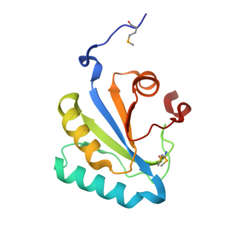 | |
UniProt | |||||
Entity Groups | |||||
| Sequence Clusters | 30% Identity50% Identity70% Identity90% Identity95% Identity100% Identity | ||||
| UniProt Group | Q13JM0 | ||||
Sequence AnnotationsExpand | |||||
Reference Sequence | |||||
| Ligands 4 Unique | |||||
|---|---|---|---|---|---|
| ID | Chains | Name / Formula / InChI Key | 2D Diagram | 3D Interactions | |
| CIT Download:Ideal Coordinates CCD File | M [auth C] | CITRIC ACID C6 H8 O7 KRKNYBCHXYNGOX-UHFFFAOYSA-N |  | ||
| PO4 Download:Ideal Coordinates CCD File | F [auth A], K [auth B], L [auth C], Q [auth D] | PHOSPHATE ION O4 P NBIIXXVUZAFLBC-UHFFFAOYSA-K |  | ||
| EDO Download:Ideal Coordinates CCD File | G [auth A], H [auth A], N [auth C], O [auth C], R [auth D] | 1,2-ETHANEDIOL C2 H6 O2 LYCAIKOWRPUZTN-UHFFFAOYSA-N |  | ||
| CL Download:Ideal Coordinates CCD File | E [auth A], I [auth B], J [auth B], P [auth D] | CHLORIDE ION Cl VEXZGXHMUGYJMC-UHFFFAOYSA-M |  | ||
| Modified Residues 1 Unique | |||||
|---|---|---|---|---|---|
| ID | Chains | Type | Formula | 2D Diagram | Parent |
| MSE Query on MSE | A, B, C, D | L-PEPTIDE LINKING | C5 H11 N O2 Se |  | MET |
| Length ( Å ) | Angle ( ˚ ) |
|---|---|
| a = 43.835 | α = 90 |
| b = 138.772 | β = 98.43 |
| c = 44.046 | γ = 90 |
| Software Name | Purpose |
|---|---|
| MolProbity | model building |
| REFMAC | refinement |
| SCALA | data scaling |
| PDB_EXTRACT | data extraction |
| MAR345 | data collection |
| MOSFLM | data reduction |
| CCP4 | data scaling |
| MOLREP | phasing |


