2P3A | pdb_00002p3a

Crystal Structure of the multi-drug resistant mutant subtype B HIV protease complexed with TL-3 inhibitor


Experimental Data Snapshot

  • Method: X-RAY DIFFRACTION
  • Resolution: 1.75 Å
  • R-Value Free: 
    0.232 (Depositor), 0.230 (DCC) 
  • R-Value Work: 
    0.183 (Depositor), 0.180 (DCC) 
  • R-Value Observed: 
    0.185 (Depositor) 

Starting Model: experimental
View more details

wwPDB Validation   3D Report Full Report


Ligand Structure Quality Assessment 

Created with Raphaël 2.3.0Worse 01 BetterLigand structure goodness of fit to experimental dataBest fitted 3TLClick on this verticalbar to view details

This is version 1.6 of the entry. See complete history


Literature

Structural Characterization of B and non-B Subtypes of HIV-Protease: Insights into the Natural Susceptibility to Drug Resistance Development.

Sanches, M.Krauchenco, S.Martins, N.H.Gustchina, A.Wlodawer, A.Polikarpov, I.

(2007) J Mol Biology 369: 1029-1040

  • DOI: https://doi.org/10.1016/j.jmb.2007.03.049
  • Primary Citation of Related Structures:  
    2P3A, 2P3B, 2P3C, 2P3D

  • PubMed Abstract: 

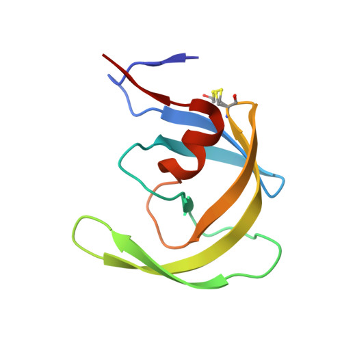
    Although a majority of HIV-1 infections in Brazil are caused by the subtype B virus (also prevalent in the United States and Western Europe), viral subtypes F and C are also found very frequently. Genomic differences between the subtypes give rise to sequence variations in the encoded proteins, including the HIV-1 protease. The current anti-HIV drugs have been developed primarily against subtype B and the effects arising from the combination of drug-resistance mutations with the naturally existing polymorphisms in non-B HIV-1 subtypes are only beginning to be elucidated. To gain more insights into the structure and function of different variants of HIV proteases, we have determined a 2.1 A structure of the native subtype F HIV-1 protease (PR) in complex with the protease inhibitor TL-3. We have also solved crystal structures of two multi-drug resistant mutant HIV PRs in complex with TL-3, from subtype B (Bmut) carrying the primary mutations V82A and L90M, and from subtype F (Fmut) carrying the primary mutation V82A plus the secondary mutation M36I, at 1.75 A and 2.8 A resolution, respectively. The proteases Bmut, Fwt and Fmut exhibit sevenfold, threefold, and 54-fold resistance to TL-3, respectively. In addition, the structure of subtype B wild type HIV-PR in complex with TL-3 has been redetermined in space group P6(1), consistent with the other three structures. Our results show that the primary mutation V82A causes the known effect of collapsing the S1/S1' pockets that ultimately lead to the reduced inhibitory effect of TL-3. Our results further indicate that two naturally occurring polymorphic substitutions in subtype F and other non-B HIV proteases, M36I and L89M, may lead to early development of drug resistance in patients infected with non-B HIV subtypes.


  • Organizational Affiliation

    Grupo de Cristalografia, Instituto de Física de São Carlos, Universidade de São Paulo, Av. Trabalhador Saocarlense, 400, CEP 13560-970, São Carlos, SP, Brazil.


Macromolecules
Find similar proteins by:  (by identity cutoff)  |  3D Structure
Entity ID: 1
MoleculeChains Sequence LengthOrganismDetailsImage
protease
A, B
99Human immunodeficiency virus 1Mutation(s): 0 
Gene Names: pol
EC: 3.4.23.16
UniProt
Find proteins for Q6BB74 (Human immunodeficiency virus type 1)
Explore Q6BB74 
Go to UniProtKB:  Q6BB74
Entity Groups  
Sequence Clusters30% Identity50% Identity70% Identity90% Identity95% Identity100% Identity
UniProt GroupQ6BB74
Sequence Annotations
Expand
  • Reference Sequence
Small Molecules
Ligands 1 Unique
IDChains Name / Formula / InChI Key2D Diagram3D Interactions
3TL
Query on 3TL

Download Ideal Coordinates CCD File 
C [auth A]benzyl [(1S,4S,7S,8R,9R,10S,13S,16S)-7,10-dibenzyl-8,9-dihydroxy-1,16-dimethyl-4,13-bis(1-methylethyl)-2,5,12,15,18-pentaoxo-20-phenyl-19-oxa-3,6,11,14,17-pentaazaicos-1-yl]carbamate
C50 H64 N6 O10
BJJPNOGMLLUCER-KUTQPOQPSA-N
Modified Residues  1 Unique
IDChains TypeFormula2D DiagramParent
CME
Query on CME
A, B
L-PEPTIDE LINKINGC5 H11 N O3 S2CYS
Biologically Interesting Molecules (External Reference) 1 Unique
Experimental Data & Validation

Experimental Data

  • Method: X-RAY DIFFRACTION
  • Resolution: 1.75 Å
  • R-Value Free:  0.232 (Depositor), 0.230 (DCC) 
  • R-Value Work:  0.183 (Depositor), 0.180 (DCC) 
  • R-Value Observed: 0.185 (Depositor) 
Space Group: P 61
Unit Cell:
Length ( Å )Angle ( ˚ )
a = 60.931α = 90
b = 60.931β = 90
c = 82.463γ = 120
Software Package:
Software NamePurpose
REFMACrefinement
PDB_EXTRACTdata extraction
MAR345data collection
MOSFLMdata reduction
CCP4data scaling
PHASERphasing

Structure Validation

View Full Validation Report


Ligand Structure Quality Assessment 

Created with Raphaël 2.3.0Worse 01 BetterLigand structure goodness of fit to experimental dataBest fitted 3TLClick on this verticalbar to view details

Entry History 

Deposition Data

Revision History  (Full details and data files)

  • Version 1.0: 2007-04-24
    Type: Initial release
  • Version 1.1: 2008-05-01
    Changes: Version format compliance
  • Version 1.2: 2011-07-13
    Changes: Atomic model, Database references, Derived calculations, Non-polymer description, Structure summary, Version format compliance
  • Version 1.3: 2013-02-27
    Changes: Other
  • Version 1.4: 2017-10-18
    Changes: Advisory, Refinement description
  • Version 1.5: 2024-04-03
    Changes: Advisory, Data collection, Database references, Derived calculations, Refinement description
  • Version 1.6: 2024-10-30
    Changes: Structure summary