2OVW | pdb_00002ovw

ENDOGLUCANASE I COMPLEXED WITH CELLOBIOSE


Experimental Data Snapshot

  • Method: X-RAY DIFFRACTION
  • Resolution: 2.30 Å
  • R-Value Work: 
    0.209 (Depositor), 0.195 (DCC) 

Starting Model: experimental
View more details

wwPDB Validation 3D Report Full Report

Validation slider image for 2OVW

Ligand Structure Quality Assessment 


This is version 3.2 of the entry. See complete history

Literature

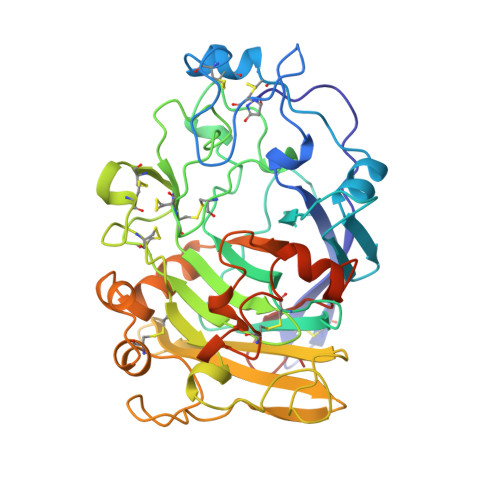
Structure of the endoglucanase I from Fusarium oxysporum: native, cellobiose, and 3,4-epoxybutyl beta-D-cellobioside-inhibited forms, at 2.3 A resolution.

Sulzenbacher, G.Schulein, M.Davies, G.J.

(1997) Biochemistry 36: 5902-5911

  • DOI: https://doi.org/10.1021/bi962963+
  • Primary Citation Related Structures: 
    2OVW, 3OVW, 4OVW

  • PubMed Abstract: 

    The mechanisms involved in the enzymatic degradation of cellulose are of great ecological and commercial importance. The breakdown of cellulose by fungal species is performed by a consortium of free enzymes, known as cellobiohydrolases and endoglucanases, which are found in many of the 57 glycosyl hydrolase families. The structure of the endoglucanase I (EG I), found in glycosyl hydrolase family 7, from the thermophilic fungus Fusarium oxysporum has been solved at 2.3 A resolution. In addition to the native enzyme, structures have also been determined with both the affinity label, 3,4-epoxybutyl beta-D-cellobioside, and the reaction product cellobiose. The affinity label is covalently bound, as expected, to the catalytic nucleophile, Glu197, with clear evidence for binding of both the R and S stereoisomers. Cellobiose is found bound to the -2 and -1 subsites of the enzyme. In marked contrast to the structure of EG I with a nonhydrolyzable thiosaccharide analog, which spanned the -2, -1, and +1 subsites and which had a skew-boat conformation for the -1 subsite sugar [Sulzenbacher, G., et al. (1996) Biochemistry 35, 15280-15287], the cellobiose complex shows no pyranoside ring distortion in the -1 subsite, implying that strain is induced primarily by the additional +1 subsite interactions and that the product is found, as expected, in its unstrained conformation.


  • Organizational Affiliation
    • Department of Chemistry, University of York, Heslington, U.K.

Macromolecule Content 

  • Total Structure Weight: 181.88 kDa 
  • Atom Count: 13,393 
  • Modeled Residue Count: 1,592 
  • Deposited Residue Count: 1,644 
  • Unique protein chains: 1

Macromolecules

Find similar proteins by:|  3D Structure
Entity ID: 1
MoleculeChains  Sequence LengthOrganismDetailsImage
ENDOGLUCANASE I
A, B, C, D
411Fusarium oxysporumMutation(s): 0 
EC: 3.2.1.4
UniProt
Find proteins for P46237 (Fusarium oxysporum)
Explore P46237 
Go to UniProtKB:  P46237
Entity Groups
Sequence Clusters30% Identity50% Identity70% Identity90% Identity95% Identity100% Identity
UniProt GroupP46237
Glycosylation
Glycosylation Sites: 2
Sequence Annotations
Expand
Reference Sequence

Oligosaccharides

Help  
Entity ID: 2
MoleculeChains Length2D Diagram GlycosylationD Interactions
beta-D-glucopyranose-(1-4)-beta-D-glucopyranose
E, F, G, H
2N/A
Glycosylation Resources
GlyTouCan: G84824ZO
GlyCosmos: G84824ZO

Small Molecules

Modified Residues  1 Unique
IDChains TypeFormula2D DiagramParent
PCA
Query on PCA
A, B, C, D
L-PEPTIDE LINKINGC5 H7 N O3GLN

Experimental Data & Validation

Experimental Data

  • Method: X-RAY DIFFRACTION
  • Resolution: 2.30 Å
  • R-Value Work:  0.209 (Depositor), 0.195 (DCC) 
Space Group: P 1 21 1
Unit Cell:
Length ( Å )Angle ( ˚ )
a = 67.93α = 90
b = 78.31β = 96.98
c = 142.47γ = 90
Software Package:
Software NamePurpose
DENZOdata reduction
SCALEPACKdata scaling
AMoREphasing
REFMACrefinement

Structure Validation

View Full Validation Report


Ligand Structure Quality Assessment 


Entry History 

Deposition Data

Revision History  (Full details and data files)

  • Version 1.0: 1998-04-08
    Type: Initial release
  • Version 1.1: 2008-03-03
    Changes: Version format compliance
  • Version 1.2: 2011-07-13
    Changes: Non-polymer description, Version format compliance
  • Version 2.0: 2019-12-25
    Changes: Derived calculations, Polymer sequence
  • Version 3.0: 2020-07-29
    Type: Remediation
    Reason: Carbohydrate remediation
    Changes: Atomic model, Data collection, Derived calculations, Non-polymer description, Structure summary
  • Version 3.1: 2024-04-03
    Changes: Data collection, Database references, Refinement description, Structure summary
  • Version 3.2: 2024-10-16
    Changes: Structure summary