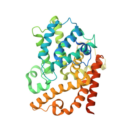
2OVV | pdb_00002ovv

Crystal structure of the catalytic domain of rat phosphodiesterase 10A


Experimental Data Snapshot

  • Method: X-RAY DIFFRACTION
  • Resolution: 2.00 Å
  • R-Value Free: 
    0.206 (Depositor), 0.214 (DCC) 
  • R-Value Work: 
    0.174 (Depositor), 0.185 (DCC) 
  • R-Value Observed: 
    0.177 (Depositor) 

wwPDB Validation   3D Report Full Report


Ligand Structure Quality Assessment 

Created with Raphaël 2.3.0Worse 01 BetterLigand structure goodness of fit to experimental dataBest fitted PFHClick on this verticalbar to view details

This is version 1.3 of the entry. See complete history


Literature

Discovery of a series of 6,7-dimethoxy-4-pyrrolidylquinazoline PDE10A inhibitors

Chappie, T.A.Humphrey, J.M.Allen, M.P.Estep, K.G.Fox, C.B.Lebel, L.A.Liras, S.Marr, E.S.Menniti, F.S.Pandit, J.Schmidt, C.J.Tu, M.Williams, R.D.Yang, F.V.

(2007) J Med Chem 50: 182-185

  • DOI: https://doi.org/10.1021/jm060653b
  • Primary Citation of Related Structures:  
    2O8H, 2OVV, 2OVY

  • PubMed Abstract: 

    A papaverine based pharmacophore model for PDE10A inhibition was generated via SBDD and used to design a library of 4-amino-6,7-dimethoxyquinazolines. From this library emerged an aryl ether pyrrolidyl 6,7-dimethoxyquinazoline series that became the focal point for additional modeling, X-ray, and synthetic efforts toward increasing PDE10A inhibitory potency and selectivity versus PDE3A/B. These efforts culminated in the discovery of 29, a potent and selective brain penetrable inhibitor of PDE10A.


  • Organizational Affiliation

    CNS Discovery and Experimental Medicinal Sciences, Pfizer Global Research and Development, Eastern Point Road, Groton, Connecticut 06340, USA. thomas.a.chappie@pfizer.com


Macromolecules
Find similar proteins by:  (by identity cutoff)  |  3D Structure
Entity ID: 1
MoleculeChains Sequence LengthOrganismDetailsImage
Phosphodiesterase-10A362Rattus norvegicusMutation(s): 0 
Gene Names: Pde10aPDE10A
EC: 3.1.4.17
UniProt
Find proteins for Q9QYJ6 (Rattus norvegicus)
Explore Q9QYJ6 
Go to UniProtKB:  Q9QYJ6
Entity Groups  
Sequence Clusters30% Identity50% Identity70% Identity90% Identity95% Identity100% Identity
UniProt GroupQ9QYJ6
Sequence Annotations
Expand
  • Reference Sequence
Experimental Data & Validation

Experimental Data

  • Method: X-RAY DIFFRACTION
  • Resolution: 2.00 Å
  • R-Value Free:  0.206 (Depositor), 0.214 (DCC) 
  • R-Value Work:  0.174 (Depositor), 0.185 (DCC) 
  • R-Value Observed: 0.177 (Depositor) 
Space Group: H 3
Unit Cell:
Length ( Å )Angle ( ˚ )
a = 120.657α = 90
b = 120.657β = 90
c = 83.273γ = 120
Software Package:
Software NamePurpose
d*TREKdata scaling
REFMACrefinement
PDB_EXTRACTdata extraction

Structure Validation

View Full Validation Report


Ligand Structure Quality Assessment 

Created with Raphaël 2.3.0Worse 01 BetterLigand structure goodness of fit to experimental dataBest fitted PFHClick on this verticalbar to view details

Entry History 

Deposition Data

Revision History  (Full details and data files)

  • Version 1.0: 2007-03-06
    Type: Initial release
  • Version 1.1: 2008-05-01
    Changes: Version format compliance
  • Version 1.2: 2011-07-13
    Changes: Derived calculations, Version format compliance
  • Version 1.3: 2024-12-25
    Changes: Advisory, Data collection, Database references, Derived calculations, Structure summary