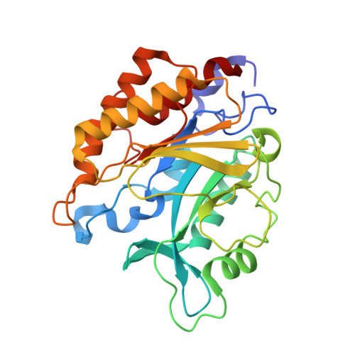
Dimer structure of an interfacially impaired phosphatidylinositol-specific phospholipase C.
Shao, C., Shi, X., Wehbi, H., Zambonelli, C., Head, J.F., Seaton, B.A., Roberts, M.F.(2007) J Biological Chem 282: 9228-9235
- PubMed: 17213187 Search on PubMed
- DOI: https://doi.org/10.1074/jbc.M610918200
- Primary Citation Related Structures:
2OR2 - PubMed Abstract:
The crystal structure of the W47A/W242A mutant of phosphatidylinositol-specific phospholipase C (PI-PLC) from Bacillus thuringiensis has been solved to 1.8A resolution. The W47A/W242A mutant is an interfacially challenged enzyme, and it has been proposed that one or both tryptophan side chains serve as membrane interfacial anchors (Feng, J., Wehbi, H., and Roberts, M. F. (2002) J. Biol. Chem. 277, 19867-19875). The crystal structure supports this hypothesis. Relative to the crystal structure of the closely related (97% identity) wild-type PI-PLC from Bacillus cereus, significant conformational differences occur at the membrane-binding interfacial region rather than the active site. The Trp --> Ala mutations not only remove the membrane-partitioning aromatic side chains but also perturb the conformations of the so-called helix B and rim loop regions, both of which are implicated in interfacial binding. The crystal structure also reveals a homodimer, the first such observation for a bacterial PI-PLC, with pseudo-2-fold symmetry. The symmetric dimer interface is stabilized by hydrophobic and hydrogen-bonding interactions, contributed primarily by a central swath of aromatic residues arranged in a quasiherringbone pattern. Evidence that interfacially active wild-type PI-PLC enzymes may dimerize in the presence of phosphatidylcholine vesicles is provided by fluorescence quenching of PI-PLC mutants with pyrene-labeled cysteine residues. The combined data suggest that wild-type PI-PLC can form similar homodimers, anchored to the interface by the tryptophan and neighboring membrane-partitioning residues.
- Boston College, Chestnut Hill, Massachusetts 02467, USA.
Organizational Affiliation: