2OJR | pdb_00002ojr

Structure of ubiquitin solved by SAD using the Lanthanide-Binding Tag


Experimental Data Snapshot

  • Method: X-RAY DIFFRACTION
  • Resolution: 2.60 Å
  • R-Value Free: 
    0.254 (Depositor), 0.260 (DCC) 
  • R-Value Work: 
    0.216 (Depositor), 0.210 (DCC) 
  • R-Value Observed: 
    0.218 (Depositor) 

wwPDB Validation 3D Report Full Report

Validation slider image for 2OJR

This is version 1.4 of the entry. See complete history

Literature

Double-Lanthanide-Binding Tags for Macromolecular Crystallographic Structure Determination.

Silvaggi, N.R.Martin, L.J.Schwalbe, H.Imperiali, B.Allen, K.N.

(2007) J Am Chem Soc 129: 7114-7120

  • DOI: https://doi.org/10.1021/ja070481n
  • Primary Citation Related Structures: 
    2OJR

  • PubMed Abstract: 

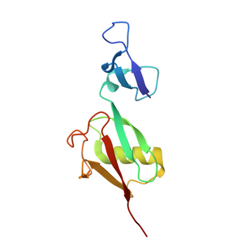
    A double-lanthanide-binding tag (dLBT), a small peptide sequence engineered to bind two lanthanide ions (e.g., Tb3+) with high affinity, was used to solve the phase problem for the structure determination of ubiquitin by the single-wavelength anomalous diffraction (SAD) method. Since the dLBT is comprised exclusively of encoded amino acids, the necessity for the incorporation of unnatural amino acids or chemical modification of the protein as a prerequisite for X-ray structure determination is eliminated. A construct encoding the dLBT as an N-terminal fusion with ubiquitin provides for facile expression and purification using standard methods. Phasing of the single-wavelength X-ray data (at 2.6 A resolution) using only the anomalous signal from the two tightly bound Tb3+ ions in the dLBT led to clear electron-density maps. Nearly 75% of the ubiquitin structure was built using automated model-building software without user intervention. It is anticipated that this technique will be broadly applicable, complementing existing macromolecular phasing methodologies. The dLBT should be particularly useful in cases where protein derivatization with heavy atoms proves to be problematic or synchrotron facilities are unavailable.


  • Organizational Affiliation
    • Department of Physiology and Biophysics, Boston University School of Medicine, 715 Albany Street, Boston, Massachusetts 02118, USA.

Macromolecule Content 

  • Total Structure Weight: 12.76 kDa 
  • Atom Count: 890 
  • Modeled Residue Count: 110 
  • Deposited Residue Count: 111 
  • Unique protein chains: 1

Macromolecules

Find similar proteins by:|  3D Structure
Entity ID: 1
MoleculeChains  Sequence LengthOrganismDetailsImage
Ubiquitin111Homo sapiensMutation(s): 0 
UniProt & NIH Common Fund Data Resources
Find proteins for P0CG48 (Homo sapiens)
Explore P0CG48 
Go to UniProtKB:  P0CG48
PHAROS:  P0CG48
GTEx:  ENSG00000150991 
Entity Groups
Sequence Clusters30% Identity50% Identity70% Identity90% Identity95% Identity100% Identity
UniProt GroupP0CG48
Sequence Annotations
Expand
Reference Sequence

Experimental Data & Validation

Experimental Data

  • Method: X-RAY DIFFRACTION
  • Resolution: 2.60 Å
  • R-Value Free:  0.254 (Depositor), 0.260 (DCC) 
  • R-Value Work:  0.216 (Depositor), 0.210 (DCC) 
  • R-Value Observed: 0.218 (Depositor) 
Space Group: P 32 2 1
Unit Cell:
Length ( Å )Angle ( ˚ )
a = 57.87α = 90
b = 57.87β = 90
c = 115.26γ = 120
Software Package:
Software NamePurpose
DENZOdata reduction
SCALEPACKdata scaling
SnBphasing
RESOLVEphasing
REFMACrefinement
PDB_EXTRACTdata extraction

Structure Validation

View Full Validation Report


Entry History 

Deposition Data

Revision History  (Full details and data files)

  • Version 1.0: 2007-06-12
    Type: Initial release
  • Version 1.1: 2008-05-01
    Changes: Version format compliance
  • Version 1.2: 2011-07-13
    Changes: Version format compliance
  • Version 1.3: 2017-10-18
    Changes: Refinement description
  • Version 1.4: 2023-12-27
    Changes: Data collection, Database references, Derived calculations