2OG7 | pdb_00002og7

Cystal structure of asparagine oxygenase in complex with Fe(II), 2S,3S-3-hydroxyasparagine and succinate


Experimental Data Snapshot

  • Method: X-RAY DIFFRACTION
  • Resolution: 1.66 Å
  • R-Value Free: 
    0.184 (Depositor), 0.200 (DCC) 
  • R-Value Work: 
    0.168 (Depositor), 0.183 (DCC) 
  • R-Value Observed: 
    0.168 (Depositor) 

Starting Model: experimental
View more details

wwPDB Validation 3D Report Full Report

Validation slider image for 2OG7

This is version 1.4 of the entry. See complete history

Literature

Mechanistic and structural basis of stereospecific Cbeta-hydroxylation in calcium-dependent antibiotic, a daptomycin-type lipopeptide

Strieker, M.Kopp, F.Mahlert, C.Essen, L.-O.Marahiel, M.A.

(2007) ACS Chem Biol 2: 187-196

  • DOI: https://doi.org/10.1021/cb700012y
  • Primary Citation Related Structures: 
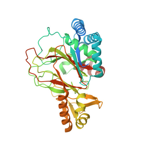
    2OG5, 2OG6, 2OG7

  • PubMed Abstract: 

    Non-ribosomally synthesized lipopeptide antibiotics of the daptomycin type are known to contain unnatural beta-modified amino acids, which are essential for bioactivity. Here we present the biochemical and structural basis for the incorporation of 3-hydroxyasparagine at position 9 in the 11-residue acidic lipopeptide lactone calcium-dependent antibiotic (CDA). Direct hydroxylation of l-asparagine by AsnO, a non-heme Fe(2+)/alpha-ketoglutarate-dependent oxygenase encoded by the CDA biosynthesis gene cluster, was validated by Fmoc derivatization of the reaction product and LC/MS analysis. The 1.45, 1.92, and 1.66 A crystal structures of AsnO as apoprotein, Fe(2+) complex, and product complex, respectively, with (2S,3S)-3-hydroxyasparagine and succinate revealed the stereoselectivity and substrate specificity of AsnO. The comparison of native and product-complex structures of AsnO showed a lid-like region (residues F208-E223) that seals the active site upon substrate binding and shields it from sterically demanding peptide substrates. Accordingly, beta-hydroxylated asparagine is synthesized prior to its incorporation into the growing CDA peptide. The AsnO structure could serve as a template for engineering novel enzymes for the synthesis of beta-hydroxylated amino acids.


  • Organizational Affiliation
    • Department of Chemistry/Biochemistry, Philipps University of Marburg, Marburg, Germany.

Macromolecule Content 

  • Total Structure Weight: 39.27 kDa 
  • Atom Count: 2,725 
  • Modeled Residue Count: 319 
  • Deposited Residue Count: 357 
  • Unique protein chains: 1

Macromolecules

Find similar proteins by:|  3D Structure
Entity ID: 1
MoleculeChains  Sequence LengthOrganismDetailsImage
asparagine oxygenase357Streptomyces coelicolor A3(2)Mutation(s): 0 
Gene Names: sco3236 (asnO)
EC: 1.14.11.39
UniProt
Find proteins for Q9Z4Z5 (Streptomyces coelicolor (strain ATCC BAA-471 / A3(2) / M145))
Explore Q9Z4Z5 
Go to UniProtKB:  Q9Z4Z5
Entity Groups
Sequence Clusters30% Identity50% Identity70% Identity90% Identity95% Identity100% Identity
UniProt GroupQ9Z4Z5
Sequence Annotations
Expand
Reference Sequence

Experimental Data & Validation

Experimental Data

  • Method: X-RAY DIFFRACTION
  • Resolution: 1.66 Å
  • R-Value Free:  0.184 (Depositor), 0.200 (DCC) 
  • R-Value Work:  0.168 (Depositor), 0.183 (DCC) 
  • R-Value Observed: 0.168 (Depositor) 
Space Group: P 31 2 1
Unit Cell:
Length ( Å )Angle ( ˚ )
a = 91.308α = 90
b = 91.308β = 90
c = 90.867γ = 120
Software Package:
Software NamePurpose
REFMACrefinement
PDB_EXTRACTdata extraction
MAR345data collection
MOSFLMdata reduction
CCP4data scaling
MOLREPphasing

Structure Validation

View Full Validation Report


Entry History 

Deposition Data

Revision History  (Full details and data files)

  • Version 1.0: 2007-03-06
    Type: Initial release
  • Version 1.1: 2008-05-01
    Changes: Version format compliance
  • Version 1.2: 2011-07-13
    Changes: Advisory, Version format compliance
  • Version 1.3: 2017-10-18
    Changes: Refinement description
  • Version 1.4: 2023-08-30
    Changes: Data collection, Database references, Derived calculations, Refinement description