2ODY | pdb_00002ody

Thrombin-bound boophilin displays a functional and accessible reactive-site loop


Experimental Data Snapshot

  • Method: X-RAY DIFFRACTION
  • Resolution: 2.35 Å
  • R-Value Free: 
    0.230 (Depositor), 0.249 (DCC) 
  • R-Value Work: 
    0.190 (Depositor) 
  • R-Value Observed: 
    0.192 (Depositor) 

Starting Model: experimental
View more details

wwPDB Validation 3D Report Full Report

Validation slider image for 2ODY

Ligand Structure Quality Assessment 


This is version 1.4 of the entry. See complete history

Literature

Isolation, cloning and structural characterisation of boophilin, a multifunctional kunitz-type proteinase inhibitor from the cattle tick.

Macedo-Ribeiro, S.Almeida, C.Calisto, B.M.Friedrich, T.Mentele, R.Sturzebecher, J.Fuentes-Prior, P.Pereira, P.J.

(2008) PLoS One 3: e1624-e1624

  • DOI: https://doi.org/10.1371/journal.pone.0001624
  • Primary Citation Related Structures: 
    2ODY

  • PubMed Abstract: 

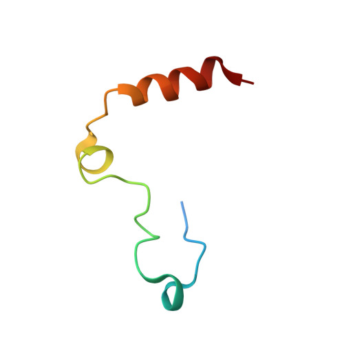
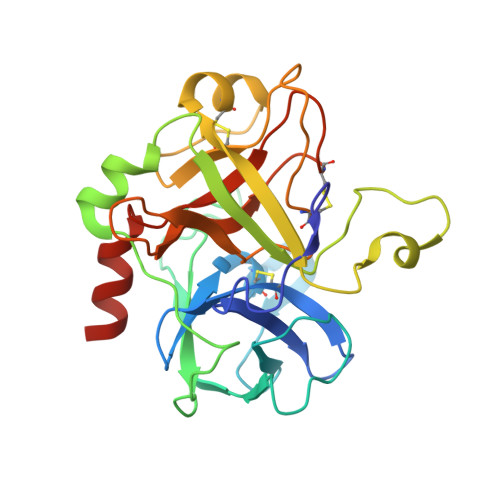
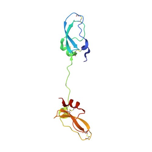
    Inhibitors of coagulation factors from blood-feeding animals display a wide variety of structural motifs and inhibition mechanisms. We have isolated a novel inhibitor from the cattle tick Boophilus microplus, one of the most widespread parasites of farm animals. The inhibitor, which we have termed boophilin, has been cloned and overexpressed in Escherichia coli. Mature boophilin is composed of two canonical Kunitz-type domains, and inhibits not only the major procoagulant enzyme, thrombin, but in addition, and by contrast to all other previously characterised natural thrombin inhibitors, significantly interferes with the proteolytic activity of other serine proteinases such as trypsin and plasmin. The crystal structure of the bovine alpha-thrombin.boophilin complex, refined at 2.35 A resolution reveals a non-canonical binding mode to the proteinase. The N-terminal region of the mature inhibitor, Q16-R17-N18, binds in a parallel manner across the active site of the proteinase, with the guanidinium group of R17 anchored in the S(1) pocket, while the C-terminal Kunitz domain is negatively charged and docks into the basic exosite I of thrombin. This binding mode resembles the previously characterised thrombin inhibitor, ornithodorin which, unlike boophilin, is composed of two distorted Kunitz modules. Unexpectedly, both boophilin domains adopt markedly different orientations when compared to those of ornithodorin, in its complex with thrombin. The N-terminal boophilin domain rotates 9 degrees and is displaced by 6 A, while the C-terminal domain rotates almost 6 degrees accompanied by a 3 A displacement. The reactive-site loop of the N-terminal Kunitz domain of boophilin with its P(1) residue, K31, is fully solvent exposed and could thus bind a second trypsin-like proteinase without sterical restraints. This finding explains the formation of a ternary thrombin.boophilin.trypsin complex, and suggests a mechanism for prothrombinase inhibition in vivo.


  • Organizational Affiliation
    • Centro de Neurociências e Biologia Celular (CNC), Coimbra, Portugal.

Macromolecule Content 

  • Total Structure Weight: 99.67 kDa 
  • Atom Count: 7,258 
  • Modeled Residue Count: 848 
  • Deposited Residue Count: 870 
  • Unique protein chains: 3

Macromolecules

Find similar proteins by:|  3D Structure
Entity ID: 1
MoleculeChains  Sequence LengthOrganismDetailsImage
Prothrombin (EC 3.4.21.5)
A, C
49Bos taurusMutation(s): 0 
EC: 3.4.21.5
UniProt
Find proteins for P00735 (Bos taurus)
Explore P00735 
Go to UniProtKB:  P00735
Entity Groups
Sequence Clusters30% Identity50% Identity70% Identity90% Identity95% Identity100% Identity
UniProt GroupP00735
Sequence Annotations
Expand
Reference Sequence
Find similar proteins by:|  3D Structure
Entity ID: 2
MoleculeChains  Sequence LengthOrganismDetailsImage
Prothrombin (EC 3.4.21.5)
B, D
259Bos taurusMutation(s): 0 
EC: 3.4.21.5
UniProt
Find proteins for P00735 (Bos taurus)
Explore P00735 
Go to UniProtKB:  P00735
Entity Groups
Sequence Clusters30% Identity50% Identity70% Identity90% Identity95% Identity100% Identity
UniProt GroupP00735
Sequence Annotations
Expand
Reference Sequence
Find similar proteins by:|  3D Structure
Entity ID: 3
MoleculeChains  Sequence LengthOrganismDetailsImage
Boophilin
E, F
127Rhipicephalus microplusMutation(s): 0 
UniProt
Find proteins for Q8WPI2 (Rhipicephalus microplus)
Explore Q8WPI2 
Go to UniProtKB:  Q8WPI2
Entity Groups
Sequence Clusters30% Identity50% Identity70% Identity90% Identity95% Identity100% Identity
UniProt GroupQ8WPI2
Sequence Annotations
Expand
Reference Sequence

Small Molecules

Ligands 3 Unique
IDChains Name / Formula / InChI Key2D Diagram3D Interactions
NAG

Query on NAG


Download:Ideal Coordinates CCD File
K [auth D]2-acetamido-2-deoxy-beta-D-glucopyranose
C8 H15 N O6
OVRNDRQMDRJTHS-FMDGEEDCSA-N
PO4

Query on PO4


Download:Ideal Coordinates CCD File
G [auth A],
H [auth B],
I [auth B],
M [auth F]
PHOSPHATE ION
O4 P
NBIIXXVUZAFLBC-UHFFFAOYSA-K
NA

Query on NA


Download:Ideal Coordinates CCD File
J [auth B],
L [auth D]
SODIUM ION
Na
FKNQFGJONOIPTF-UHFFFAOYSA-N

Experimental Data & Validation

Experimental Data

  • Method: X-RAY DIFFRACTION
  • Resolution: 2.35 Å
  • R-Value Free:  0.230 (Depositor), 0.249 (DCC) 
  • R-Value Work:  0.190 (Depositor) 
  • R-Value Observed: 0.192 (Depositor) 
Space Group: P 21 21 21
Unit Cell:
Length ( Å )Angle ( ˚ )
a = 92.5α = 90
b = 104.2β = 90
c = 129.1γ = 90
Software Package:
Software NamePurpose
REFMACrefinement
MOSFLMdata reduction
SCALAdata scaling
AMoREphasing

Structure Validation

View Full Validation Report


Ligand Structure Quality Assessment 


Entry History 

Deposition Data

Revision History  (Full details and data files)

  • Version 1.0: 2008-01-22
    Type: Initial release
  • Version 1.1: 2011-07-13
    Changes: Advisory, Derived calculations, Version format compliance
  • Version 1.2: 2020-07-29
    Type: Remediation
    Reason: Carbohydrate remediation
    Changes: Advisory, Data collection, Derived calculations, Structure summary
  • Version 1.3: 2023-08-30
    Changes: Advisory, Data collection, Database references, Refinement description, Structure summary
  • Version 1.4: 2024-10-30
    Changes: Structure summary