2OD3

Human thrombin chimera with human residues 184a, 186, 186a, 186b, 186c and 222 replaced by murine thrombin equivalents.


Experimental Data Snapshot

  • Method: X-RAY DIFFRACTION
  • Resolution: 1.75 Å
  • R-Value Free: 
    0.230 (Depositor), 0.220 (DCC) 
  • R-Value Work: 
    0.199 (Depositor), 0.190 (DCC) 
  • R-Value Observed: 
    0.199 (Depositor) 

Starting Model: experimental
View more details

wwPDB Validation   3D Report Full Report


Ligand Structure Quality Assessment 

Created with Raphaël 2.3.0Worse 01 BetterLigand structure goodness of fit to experimental dataBest fitted 0G6Click on this verticalbar to view detailsBest fitted NAGClick on this verticalbar to view details

This is version 1.8 of the entry. See complete history


Literature

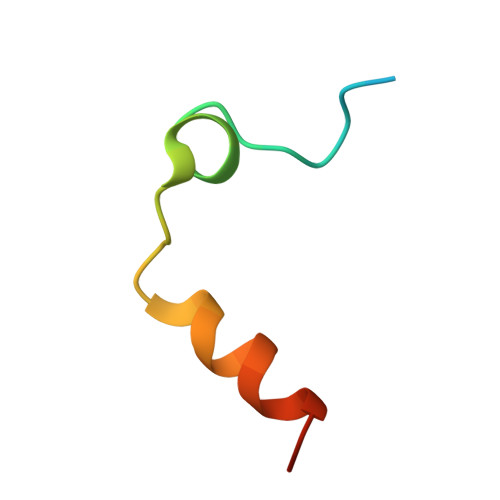
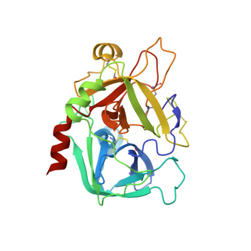
Structural basis of na+ activation mimicry in murine thrombin.

Marino, F.Chen, Z.W.Ergenekan, C.E.Bush-Pelc, L.A.Mathews, F.S.Di Cera, E.

(2007) J Biol Chem 282: 16355-16361

  • DOI: https://doi.org/10.1074/jbc.M701323200
  • Primary Citation of Related Structures:  
    2OCV, 2OD3

  • PubMed Abstract: 

    Unlike human thrombin, murine thrombin lacks Na+ activation due to the charge reversal substitution D222K in the Na+ binding loop. However, the enzyme is functionally stabilized in a Na+-bound form and is highly active toward physiologic substrates. The structural basis of this peculiar property is unknown. Here, we present the 2.2 A resolution x-ray crystal structure of murine thrombin in the absence of inhibitors and salts. The enzyme assumes an active conformation, with Ser-195, Glu-192, and Asp-189 oriented as in the Na+-bound fast form of human thrombin. Lys-222 completely occludes the pore of entry to the Na+ binding site and positions its side chain inside the pore, with the Nzeta atom H-bonded to the backbone oxygen atoms of Lys-185, Asp-186b, and Lys-186d. The same architecture is observed in the 1.75 A resolution structure of a thrombin chimera in which the human enzyme carries all residues defining the Na+ pore in the murine enzyme. These findings demonstrate that Na+ activation in thrombin is linked to the architecture of the Na+ pore. The molecular strategy of Na+ activation mimicry unraveled for murine thrombin is relevant to serine proteases and enzymes activated by monovalent cations in general.


  • Organizational Affiliation

    Department of Biochemistry and Molecular Biophysics, Washington University Medical School, St. Louis, Missouri 63110, USA.


Macromolecules
Find similar proteins by:  (by identity cutoff)  |  3D Structure
Entity ID: 1
MoleculeChains Sequence LengthOrganismDetailsImage
Thrombin light chain36Homo sapiensMutation(s): 0 
Gene Names: F2
EC: 3.4.21.5
UniProt & NIH Common Fund Data Resources
Find proteins for P00734 (Homo sapiens)
Explore P00734 
Go to UniProtKB:  P00734
PHAROS:  P00734
GTEx:  ENSG00000180210 
Entity Groups  
Sequence Clusters30% Identity50% Identity70% Identity90% Identity95% Identity100% Identity
UniProt GroupP00734
Sequence Annotations
Expand
  • Reference Sequence
Find similar proteins by:  (by identity cutoff)  |  3D Structure
Entity ID: 2
MoleculeChains Sequence LengthOrganismDetailsImage
Thrombin heavy chain259Homo sapiensMutation(s): 6 
Gene Names: F2
EC: 3.4.21.5
UniProt & NIH Common Fund Data Resources
Find proteins for P00734 (Homo sapiens)
Explore P00734 
Go to UniProtKB:  P00734
PHAROS:  P00734
GTEx:  ENSG00000180210 
Entity Groups  
Sequence Clusters30% Identity50% Identity70% Identity90% Identity95% Identity100% Identity
UniProt GroupP00734
Glycosylation
Glycosylation Sites: 1Go to GlyGen: P00734-1
Sequence Annotations
Expand
  • Reference Sequence
Small Molecules
Ligands 2 Unique
IDChains Name / Formula / InChI Key2D Diagram3D Interactions
0G6
Query on 0G6

Download Ideal Coordinates CCD File 
C [auth B]D-phenylalanyl-N-[(2S,3S)-6-{[amino(iminio)methyl]amino}-1-chloro-2-hydroxyhexan-3-yl]-L-prolinamide
C21 H34 Cl N6 O3
DVFLYEYCMMLBTQ-VSZNYVQBSA-O
NAG
Query on NAG

Download Ideal Coordinates CCD File 
D [auth B]2-acetamido-2-deoxy-beta-D-glucopyranose
C8 H15 N O6
OVRNDRQMDRJTHS-FMDGEEDCSA-N
Biologically Interesting Molecules (External Reference) 1 Unique
Experimental Data & Validation

Experimental Data

  • Method: X-RAY DIFFRACTION
  • Resolution: 1.75 Å
  • R-Value Free:  0.230 (Depositor), 0.220 (DCC) 
  • R-Value Work:  0.199 (Depositor), 0.190 (DCC) 
  • R-Value Observed: 0.199 (Depositor) 
Space Group: P 21 21 21
Unit Cell:
Length ( Å )Angle ( ˚ )
a = 44.952α = 90
b = 74.078β = 90
c = 103.565γ = 90
Software Package:
Software NamePurpose
CNSrefinement
ADSCdata collection
HKL-2000data reduction
SCALEPACKdata scaling
CCP4phasing

Structure Validation

View Full Validation Report


Ligand Structure Quality Assessment 

Created with Raphaël 2.3.0Worse 01 BetterLigand structure goodness of fit to experimental dataBest fitted 0G6Click on this verticalbar to view detailsBest fitted NAGClick on this verticalbar to view details

Entry History 

Deposition Data

Revision History  (Full details and data files)

  • Version 1.0: 2007-04-10
    Type: Initial release
  • Version 1.1: 2008-05-01
    Changes: Version format compliance
  • Version 1.2: 2011-07-13
    Changes: Atomic model, Database references, Derived calculations, Non-polymer description, Structure summary, Version format compliance
  • Version 1.3: 2013-02-27
    Changes: Other
  • Version 1.4: 2020-07-29
    Type: Remediation
    Reason: Carbohydrate remediation
    Changes: Data collection, Derived calculations, Structure summary
  • Version 1.5: 2021-10-20
    Changes: Database references, Structure summary
  • Version 1.6: 2023-08-30
    Changes: Data collection, Refinement description
  • Version 1.7: 2024-03-13
    Changes: Source and taxonomy, Structure summary
  • Version 1.8: 2024-10-16
    Changes: Structure summary