2O98 | pdb_00002o98

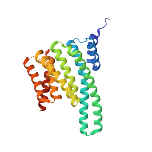
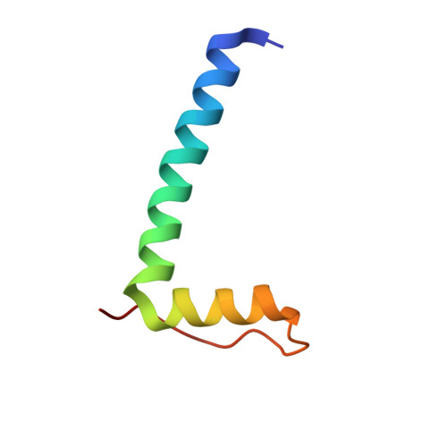
Structure of the 14-3-3 / H+-ATPase plant complex


Experimental Data Snapshot

  • Method: X-RAY DIFFRACTION
  • Resolution: 2.70 Å
  • R-Value Free: 
    0.263 (Depositor), 0.255 (DCC) 
  • R-Value Work: 
    0.178 (Depositor), 0.174 (DCC) 
  • R-Value Observed: 
    0.182 (Depositor) 

wwPDB Validation 3D Report Full Report

Validation slider image for 2O98

Ligand Structure Quality Assessment 


This is version 1.5 of the entry. See complete history. 

Literature

Structure of a 14-3-3 coordinated hexamer of the plant plasma membrane H+ -ATPase by combining X-ray crystallography and electron cryomicroscopy

Ottmann, C., Marco, S., Jaspert, N., Marcon, C., Schauer, N., Weyand, M., Vandermeeren, C., Duby, G., Boutry, M., Wittinghofer, A., Rigaud, J.L., Oecking, C.

(2007) Mol Cell 25: 427-440

  • DOI: https://doi.org/10.1016/j.molcel.2006.12.017
  • Primary Citation Related Structures: 
    2O98

  • PubMed Abstract: 

    Regulatory 14-3-3 proteins activate the plant plasma membrane H(+)-ATPase by binding to its C-terminal autoinhibitory domain. This interaction requires phosphorylation of a C-terminal, mode III, recognition motif as well as an adjacent span of approximately 50 amino acids. Here we report the X-ray crystal structure of 14-3-3 in complex with the entire binding motif, revealing a previously unidentified mode of interaction. A 14-3-3 dimer simultaneously binds two H(+)-ATPase peptides, each of which forms a loop within the typical 14-3-3 binding groove and therefore exits from the center of the dimer. Several H(+)-ATPase mutants support this structure determination. Accordingly, 14-3-3 binding could result in H(+)-ATPase oligomerization. Indeed, by using single-particle electron cryomicroscopy, the 3D reconstruction of the purified H(+)-ATPase/14-3-3 complex demonstrates a hexameric arrangement. Fitting of 14-3-3 and H(+)-ATPase atomic structures into the 3D reconstruction map suggests the spatial arrangement of the holocomplex.


  • Organizational Affiliation: 
    • Zentrum für Molekularbiologie der Pflanzen, Pflanzenphysiologie, Universität Tübingen, Auf der Morgenstelle 5, 72076 Tübingen, Germany.

Macromolecule Content 

  • Total Structure Weight: 68.5 kDa 
  • Atom Count: 4,824 
  • Modeled Residue Count: 573 
  • Deposited Residue Count: 588 
  • Unique protein chains: 2

Macromolecules

Find similar proteins by:|  3D Structure
Entity ID: 1
MoleculeChains  Sequence LengthOrganismDetailsImage
14-3-3-like protein C
A, B
242Nicotiana tabacumMutation(s): 0 
UniProt
Find proteins for P93343 (Nicotiana tabacum)
Explore P93343 
Go to UniProtKB:  P93343
Entity Groups
Sequence Clusters30% Identity50% Identity70% Identity90% Identity95% Identity100% Identity
UniProt GroupP93343
Sequence Annotations
Expand
Reference Sequence
Find similar proteins by:|  3D Structure
Entity ID: 2
MoleculeChains  Sequence LengthOrganismDetailsImage
Plasma membrane H+ ATPaseC [auth P],
D [auth Q]
52Nicotiana plumbaginifoliaMutation(s): 2 
Gene Names: pma2
EC: 7.1.2.1
UniProt
Find proteins for Q08436 (Nicotiana plumbaginifolia)
Explore Q08436 
Go to UniProtKB:  Q08436
Entity Groups
Sequence Clusters30% Identity50% Identity70% Identity90% Identity95% Identity100% Identity
UniProt GroupQ08436
Sequence Annotations
Expand
Reference Sequence

Experimental Data & Validation

Experimental Data

  • Method: X-RAY DIFFRACTION
  • Resolution: 2.70 Å
  • R-Value Free:  0.263 (Depositor), 0.255 (DCC) 
  • R-Value Work:  0.178 (Depositor), 0.174 (DCC) 
  • R-Value Observed: 0.182 (Depositor) 
Space Group: I 41 2 2
Unit Cell:
Length ( Å )Angle ( ˚ )
a = 114.37α = 90
b = 114.37β = 90
c = 236.79γ = 90
Software Package:
Software NamePurpose
REFMACrefinement
PDB_EXTRACTdata extraction
MAR345data collection
XDSdata reduction
XSCALEdata scaling
AMoREphasing

Structure Validation

View Full Validation Report


Ligand Structure Quality Assessment 


Entry History 

Deposition Data

Revision History  (Full details and data files)

  • Version 1.0: 2007-04-03
    Type: Initial release
  • Version 1.1: 2008-05-01
    Changes: Version format compliance
  • Version 1.2: 2011-07-13
    Changes: Advisory, Version format compliance
  • Version 1.3: 2017-10-18
    Changes: Advisory, Refinement description
  • Version 1.4: 2021-11-10
    Changes: Advisory, Database references, Derived calculations
  • Version 1.5: 2023-12-27
    Changes: Data collection