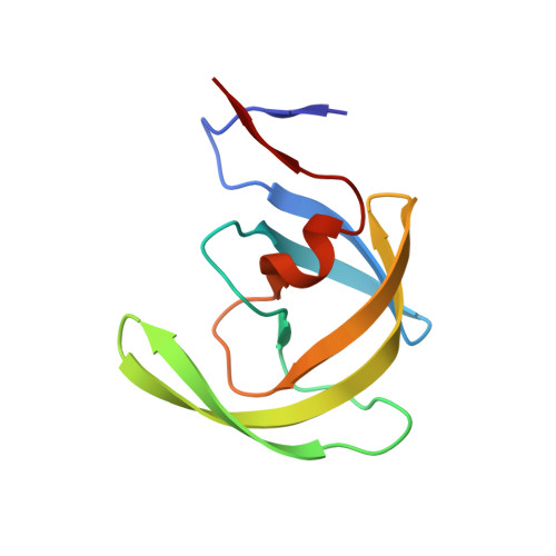
2O4K

Crystal Structure of HIV-1 Protease (Q7K) in Complex with Atazanavir


Experimental Data Snapshot

  • Method: X-RAY DIFFRACTION
  • Resolution: 1.60 Å
  • R-Value Free: 0.209 
  • R-Value Work: 0.183 
  • R-Value Observed: 0.186 

Starting Model: experimental
View more details

wwPDB Validation   3D Report Full Report


This is version 1.5 of the entry. See complete history


Literature

Unique thermodynamic response of tipranavir to human immunodeficiency virus type 1 protease drug resistance mutations.

Muzammil, S.Armstrong, A.A.Kang, L.W.Jakalian, A.Bonneau, P.R.Schmelmer, V.Amzel, L.M.Freire, E.

(2007) J Virol 81: 5144-5154

  • DOI: https://doi.org/10.1128/JVI.02706-06
  • Primary Citation of Related Structures:  
    2O4K, 2O4L, 2O4N, 2O4P, 2O4S

  • PubMed Abstract: 

    Drug resistance is a major problem affecting the clinical efficacy of antiretroviral agents, including protease inhibitors, in the treatment of infection with human immunodeficiency virus type 1 (HIV-1)/AIDS. Consequently, the elucidation of the mechanisms by which HIV-1 protease inhibitors maintain antiviral activity in the presence of mutations is critical to the development of superior inhibitors. Tipranavir, a nonpeptidic HIV-1 protease inhibitor, has been recently approved for the treatment of HIV infection. Tipranavir inhibits wild-type protease with high potency (K(i) = 19 pM) and demonstrates durable efficacy in the treatment of patients infected with HIV-1 strains containing multiple common mutations associated with resistance. The high potency of tipranavir results from a very large favorable entropy change (-TDeltaS = -14.6 kcal/mol) combined with a favorable, albeit small, enthalpy change (DeltaH = -0.7 kcal/mol, 25 degrees C). Characterization of tipranavir binding to wild-type protease, active site mutants I50V and V82F/I84V, the multidrug-resistant mutant L10I/L33I/M46I/I54V/L63I/V82A/I84V/L90M, and the tipranavir in vitro-selected mutant I13V/V32L/L33F/K45I/V82L/I84V was performed by isothermal titration calorimetry and crystallography. Thermodynamically, the good response of tipranavir arises from a unique behavior: it compensates for entropic losses by actual enthalpic gains or by sustaining minimal enthalpic losses when facing the mutants. The net result is a small loss in binding affinity. Structurally, tipranavir establishes a very strong hydrogen bond network with invariant regions of the protease, which is maintained with the mutants, including catalytic Asp25 and the backbone of Asp29, Asp30, Gly48 and Ile50. Moreover, tipranavir forms hydrogen bonds directly to Ile50, while all other inhibitors do so by being mediated by a water molecule.


  • Organizational Affiliation

    Department of Biology, Johns Hopkins University, Baltimore, MD 21218, USA.


Macromolecules
Find similar proteins by:  (by identity cutoff)  |  3D Structure
Entity ID: 1
MoleculeChains Sequence LengthOrganismDetailsImage
protease
A, B
99Human immunodeficiency virus 1Mutation(s): 1 
Gene Names: gag-pol
UniProt
Find proteins for P03367 (Human immunodeficiency virus type 1 group M subtype B (isolate BRU/LAI))
Go to UniProtKB:  P03367
Find proteins for Q903J0 (Human immunodeficiency virus 1)
Explore Q903J0 
Go to UniProtKB:  Q903J0
Entity Groups  
Sequence Clusters30% Identity50% Identity70% Identity90% Identity95% Identity100% Identity
UniProt GroupQ903J0
Sequence Annotations
Expand
  • Reference Sequence
Small Molecules
Ligands 2 Unique
IDChains Name / Formula / InChI Key2D Diagram3D Interactions
DR7
Query on DR7

Download Ideal Coordinates CCD File 
D [auth A](3S,8S,9S,12S)-3,12-BIS(1,1-DIMETHYLETHYL)-8-HYDROXY-4,11-DIOXO-9-(PHENYLMETHYL)-6-[[4-(2-PYRIDINYL)PHENYL]METHYL]-2,5, 6,10,13-PENTAAZATETRADECANEDIOIC ACID DIMETHYL ESTER
C38 H52 N6 O7
AXRYRYVKAWYZBR-GASGPIRDSA-N
CL
Query on CL

Download Ideal Coordinates CCD File 
C [auth A]CHLORIDE ION
Cl
VEXZGXHMUGYJMC-UHFFFAOYSA-M
Binding Affinity Annotations 
IDSourceBinding Affinity
DR7 BindingDB:  2O4K Ki: min: 0.05, max: 0.76 (nM) from 8 assay(s)
Kd: 0.4 (nM) from 1 assay(s)
IC50: 4 (nM) from 1 assay(s)
PDBBind:  2O4K Kd: 0.04 (nM) from 1 assay(s)
Experimental Data & Validation

Experimental Data

  • Method: X-RAY DIFFRACTION
  • Resolution: 1.60 Å
  • R-Value Free: 0.209 
  • R-Value Work: 0.183 
  • R-Value Observed: 0.186 
  • Space Group: P 21 21 2
Unit Cell:
Length ( Å )Angle ( ˚ )
a = 58.741α = 90
b = 85.805β = 90
c = 46.242γ = 90
Software Package:
Software NamePurpose
DENZOdata reduction
SCALEPACKdata scaling
MOLREPphasing
REFMACrefinement
PDB_EXTRACTdata extraction

Structure Validation

View Full Validation Report


Entry History 

Deposition Data

Revision History  (Full details and data files)

  • Version 1.0: 2006-12-12
    Type: Initial release
  • Version 1.1: 2008-04-01
    Changes: Version format compliance
  • Version 1.2: 2011-07-13
    Changes: Derived calculations, Version format compliance
  • Version 1.3: 2017-10-18
    Changes: Refinement description
  • Version 1.4: 2021-10-20
    Changes: Database references, Derived calculations, Structure summary
  • Version 1.5: 2023-08-30
    Changes: Data collection, Refinement description