2NTN

Crystal structure of MabA-C60V/G139A/S144L


Experimental Data Snapshot

  • Method: X-RAY DIFFRACTION
  • Resolution: 2.30 Å
  • R-Value Free: 0.238 
  • R-Value Work: 0.177 
  • R-Value Observed: 0.181 

Starting Model: experimental
View more details

wwPDB Validation   3D Report Full Report


This is version 1.4 of the entry. See complete history


Literature

Lack of dynamics in the MabA active site kills the enzyme activity: practical consequences for drug-design studies

Poncet-Montange, G.Ducasse-Cabanot, S.Quemard, A.Labesse, G.Cohen-Gonsaud, M.

(2007) Acta Crystallogr D Biol Crystallogr 63: 923-925

  • DOI: https://doi.org/10.1107/S0907444907024158
  • Primary Citation of Related Structures:  
    2NTN

  • PubMed Abstract: 

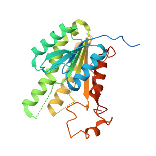
    The MabA protein from Mycobacterium tuberculosis is a validated drug target. Previous structural studies of this protein showed dynamic behaviour in the catalytic site and described motion between an open 'active' holo form (with NADP) and a closed 'inactive' apo form (without NADP). Here, a mutation (G139A) is reported that leads to complete protein inactivation and freezes the catalytic site into its closed form, even in the presence of the cofactor. This observation suggests a new way to develop anti-MabA drugs via protein stabilization of the 'inactive' form.


  • Organizational Affiliation

    CNRS UMR5048, Centre de Biochimie Structurale, F-34090 Montpellier, France.


Macromolecules
Find similar proteins by:  (by identity cutoff)  |  3D Structure
Entity ID: 1
MoleculeChains Sequence LengthOrganismDetailsImage
3-oxoacyl-[acyl-carrier-protein] reductase
A, B
267Mycobacterium tuberculosis H37RvMutation(s): 3 
Gene Names: fabGfabG1
EC: 1.1.1.100
UniProt
Find proteins for P9WGT3 (Mycobacterium tuberculosis (strain ATCC 25618 / H37Rv))
Explore P9WGT3 
Go to UniProtKB:  P9WGT3
Entity Groups  
Sequence Clusters30% Identity50% Identity70% Identity90% Identity95% Identity100% Identity
UniProt GroupP9WGT3
Sequence Annotations
Expand
  • Reference Sequence
Experimental Data & Validation

Experimental Data

  • Method: X-RAY DIFFRACTION
  • Resolution: 2.30 Å
  • R-Value Free: 0.238 
  • R-Value Work: 0.177 
  • R-Value Observed: 0.181 
  • Space Group: C 1 2 1
Unit Cell:
Length ( Å )Angle ( ˚ )
a = 81.613α = 90
b = 117.285β = 121.9
c = 52.272γ = 90
Software Package:
Software NamePurpose
REFMACrefinement
MOSFLMdata reduction
CCP4data scaling

Structure Validation

View Full Validation Report


Entry History 

Deposition Data

Revision History  (Full details and data files)

  • Version 1.0: 2006-11-21
    Type: Initial release
  • Version 1.1: 2008-05-01
    Changes: Version format compliance
  • Version 1.2: 2011-07-13
    Changes: Advisory, Derived calculations, Version format compliance
  • Version 1.3: 2021-11-10
    Changes: Database references
  • Version 1.4: 2023-10-25
    Changes: Data collection, Refinement description