2NSL | pdb_00002nsl

E. coli PurE H45N mutant complexed with CAIR


Experimental Data Snapshot

  • Method: X-RAY DIFFRACTION
  • Resolution: 2.00 Å
  • R-Value Free: 
    0.224 (Depositor), 0.222 (DCC) 
  • R-Value Work: 
    0.181 (Depositor), 0.181 (DCC) 
  • R-Value Observed: 
    0.181 (Depositor) 

Starting Model: experimental
View more details

wwPDB Validation 3D Report Full Report

Validation slider image for 2NSL

Ligand Structure Quality Assessment 


This is version 2.1 of the entry. See complete history

Literature

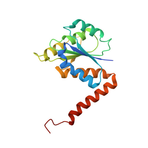
N(5)-CAIR Mutase: Role of a CO(2) Binding Site and Substrate Movement in Catalysis.

Hoskins, A.A.Morar, M.Kappock, T.J.Mathews, I.I.Zaugg, J.B.Barder, T.E.Peng, P.Okamoto, A.Ealick, S.E.Stubbe, J.

(2007) Biochemistry 46: 2842-2855

  • DOI: https://doi.org/10.1021/bi602436g
  • Primary Citation Related Structures: 
    2ATE, 2NSH, 2NSJ, 2NSL

  • PubMed Abstract: 

    N5-Carboxyaminoimidazole ribonucleotide mutase (N5-CAIR mutase or PurE) from Escherichia coli catalyzes the reversible interconversion of N5-CAIR to carboxyaminoimidazole ribonucleotide (CAIR) with direct CO2 transfer. Site-directed mutagenesis, a pH-rate profile, DFT calculations, and X-ray crystallography together provide new insight into the mechanism of this unusual transformation. These studies suggest that a conserved, protonated histidine (His45) plays an essential role in catalysis. The importance of proton transfers is supported by DFT calculations on CAIR and N5-CAIR analogues in which the ribose 5'-phosphate is replaced with a methyl group. The calculations suggest that the nonaromatic tautomer of CAIR (isoCAIR) is only 3.1 kcal/mol higher in energy than its aromatic counterpart, implicating this species as a potential intermediate in the PurE-catalyzed reaction. A structure of wild-type PurE cocrystallized with 4-nitroaminoimidazole ribonucleotide (NO2-AIR, a CAIR analogue) and structures of H45N and H45Q PurEs soaked with CAIR have been determined and provide the first insight into the binding of an intact PurE substrate. A comparison of 19 available structures of PurE and PurE mutants in apo and nucleotide-bound forms reveals a common, buried carboxylate or CO2 binding site for CAIR and N5-CAIR in a hydrophobic pocket in which the carboxylate or CO2 interacts with backbone amides. This work has led to a mechanistic proposal in which the carboxylate orients the substrate for proton transfer from His45 to N5-CAIR to form an enzyme-bound aminoimidazole ribonucleotide (AIR) and CO2 intermediate. Subsequent movement of the aminoimidazole moiety of AIR reorients it for addition of CO2 at C4 to generate isoCAIR. His45 is now in a position to remove a C4 proton to produce CAIR.


  • Organizational Affiliation
    • Department of Chemistry, Massachusetts Institute of Technology, Cambridge, Massachusetts 02139, USA.

Macromolecule Content 

  • Total Structure Weight: 18.11 kDa 
  • Atom Count: 1,340 
  • Modeled Residue Count: 162 
  • Deposited Residue Count: 169 
  • Unique protein chains: 1

Macromolecules

Find similar proteins by:|  3D Structure
Entity ID: 1
MoleculeChains  Sequence LengthOrganismDetailsImage
Phosphoribosylaminoimidazole carboxylase catalytic subunit169Escherichia coliMutation(s): 1 
Gene Names: purE
EC: 4.1.1.21 (PDB Primary Data), 5.4.99.18 (UniProt)
UniProt
Find proteins for P0AG18 (Escherichia coli (strain K12))
Explore P0AG18 
Go to UniProtKB:  P0AG18
Entity Groups
Sequence Clusters30% Identity50% Identity70% Identity90% Identity95% Identity100% Identity
UniProt GroupP0AG18
Sequence Annotations
Expand
Reference Sequence

Small Molecules

Ligands 1 Unique
IDChains Name / Formula / InChI Key2D Diagram3D Interactions
C2R

Query on C2R


Download:Ideal Coordinates CCD File
B [auth A]5-AMINO-1-(5-O-PHOSPHONO-BETA-D-RIBOFURANOSYL)-1H-IMIDAZOLE-4-CARBOXYLIC ACID
C9 H14 N3 O9 P
XFVULMDJZXYMSG-ZIYNGMLESA-N

Experimental Data & Validation

Experimental Data

  • Method: X-RAY DIFFRACTION
  • Resolution: 2.00 Å
  • R-Value Free:  0.224 (Depositor), 0.222 (DCC) 
  • R-Value Work:  0.181 (Depositor), 0.181 (DCC) 
  • R-Value Observed: 0.181 (Depositor) 
Space Group: I 4 2 2
Unit Cell:
Length ( Å )Angle ( ˚ )
a = 111.317α = 90
b = 111.317β = 90
c = 49.2γ = 90
Software Package:
Software NamePurpose
CNSrefinement
HKL-2000data collection
HKL-2000data reduction
HKL-2000data scaling
CNSphasing

Structure Validation

View Full Validation Report


Ligand Structure Quality Assessment 


Entry History 

Deposition Data

Revision History  (Full details and data files)

  • Version 1.0: 2007-04-24
    Type: Initial release
  • Version 1.1: 2008-05-01
    Changes: Version format compliance
  • Version 1.2: 2011-07-13
    Changes: Derived calculations, Version format compliance
  • Version 2.0: 2021-10-20
    Changes: Atomic model, Database references, Derived calculations, Structure summary
  • Version 2.1: 2023-08-30
    Changes: Data collection, Refinement description