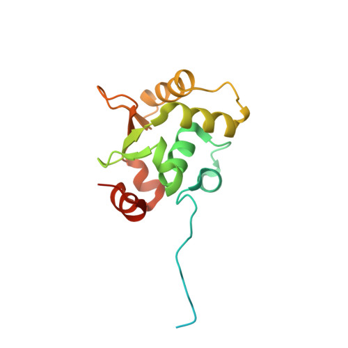
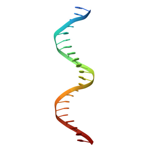
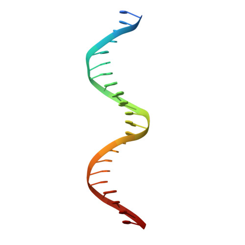
2NNY

Crystal structure of the Ets1 dimer DNA complex.


Experimental Data Snapshot

  • Method: X-RAY DIFFRACTION
  • Resolution: 2.58 Å
  • R-Value Free: 0.271 
  • R-Value Work: 0.235 

Starting Model: experimental
View more details

wwPDB Validation   3D Report Full Report


This is version 1.3 of the entry. See complete history


Literature

Regulation of the transcription factor Ets-1 by DNA-mediated homo-dimerization.

Lamber, E.P.Vanhille, L.Textor, L.C.Kachalova, G.S.Sieweke, M.H.Wilmanns, M.

(2008) EMBO J 27: 2006-2017

  • DOI: https://doi.org/10.1038/emboj.2008.117
  • Primary Citation of Related Structures:  
    2NNY

  • PubMed Abstract: 

    The function of the Ets-1 transcription factor is regulated by two regions that flank its DNA-binding domain. A previously established mechanism for auto-inhibition of monomeric Ets-1 on DNA response elements with a single ETS-binding site, however, has not been observed for the stromelysin-1 promoter containing two palindromic ETS-binding sites. We present the structure of Ets-1 on this promoter element, revealing a ternary complex in which protein homo-dimerization is mediated by the specific arrangement of the two ETS-binding sites. In this complex, the N-terminal-flanking region is required for ternary protein-DNA assembly. Ets-1 variants, in which residues from this region are mutated, loose the ability for DNA-mediated dimerization and stromelysin-1 promoter transactivation. Thus, our data unravel the molecular basis for relief of auto-inhibition and the ability of Ets-1 to function as a facultative dimeric transcription factor on this site. Our findings may also explain previous data of Ets-1 function in the context of heterologous transcription factors, thus providing a molecular model that could also be valid for Ets-1 regulation by hetero-oligomeric assembly.


  • Organizational Affiliation

    EMBL-Hamburg, c/o DESY, Hamburg, Germany.


Macromolecules

Find similar proteins by:  (by identity cutoff)  |  3D Structure
Entity ID: 3
MoleculeChains Sequence LengthOrganismDetailsImage
C-ets-1 proteinC [auth A],
D [auth B]
171Homo sapiensMutation(s): 2 
Gene Names: ETS1
UniProt & NIH Common Fund Data Resources
Find proteins for P14921 (Homo sapiens)
Explore P14921 
Go to UniProtKB:  P14921
PHAROS:  P14921
GTEx:  ENSG00000134954 
Entity Groups  
Sequence Clusters30% Identity50% Identity70% Identity90% Identity95% Identity100% Identity
UniProt GroupP14921
Sequence Annotations
Expand
  • Reference Sequence

Find similar nucleic acids by:  Sequence   |   3D Structure  

Entity ID: 1
MoleculeChains LengthOrganismImage
5'-D(*T*AP*GP*AP*CP*AP*GP*GP*AP*AP*GP*CP*AP*CP*TP*TP*CP*CP*TP*GP*GP*AP*G)-3'A [auth C]23N/A
Sequence Annotations
Expand
  • Reference Sequence

Find similar nucleic acids by:  Sequence   |   3D Structure  

Entity ID: 2
MoleculeChains LengthOrganismImage
5'-D(*A*CP*TP*CP*CP*AP*GP*GP*AP*AP*GP*TP*GP*CP*TP*TP*CP*CP*TP*GP*TP*CP*T)-3'B [auth D]23N/A
Sequence Annotations
Expand
  • Reference Sequence
Experimental Data & Validation

Experimental Data

  • Method: X-RAY DIFFRACTION
  • Resolution: 2.58 Å
  • R-Value Free: 0.271 
  • R-Value Work: 0.235 
  • Space Group: P 21 21 2
Unit Cell:
Length ( Å )Angle ( ˚ )
a = 93.556α = 90
b = 100.838β = 90
c = 69.8γ = 90
Software Package:
Software NamePurpose
ADSCdata collection
MOLREPphasing
CNSrefinement
DENZOdata reduction
SCALEPACKdata scaling

Structure Validation

View Full Validation Report


Entry History 

Deposition Data

Revision History  (Full details and data files)

  • Version 1.0: 2008-03-04
    Type: Initial release
  • Version 1.1: 2011-07-13
    Changes: Version format compliance
  • Version 1.2: 2021-10-20
    Changes: Database references
  • Version 1.3: 2023-08-30
    Changes: Data collection, Refinement description