2NMV | pdb_00002nmv

Damage detection by the UvrABC pathway: Crystal structure of UvrB bound to fluorescein-adducted DNA


Experimental Data Snapshot

  • Method: X-RAY DIFFRACTION
  • Resolution: 2.95 Å
  • R-Value Free: 
    0.281 (Depositor) 
  • R-Value Work: 
    0.224 (Depositor) 
  • R-Value Observed: 
    0.227 (Depositor) 

Starting Model: experimental
View more details

wwPDB Validation 3D Report Full Report

Validation slider image for 2NMV

This is version 1.3 of the entry. See complete history

Literature

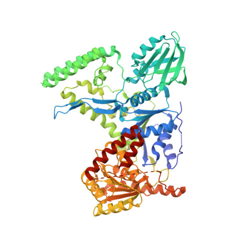
Damage detection by the UvrABC pathway: crystal structure of UvrB bound to fluorescein-adducted DNA

Waters, T.R.Eryilmaz, J.Geddes, S.Barrett, T.E.

(2006) FEBS Lett 580: 6423-6427

  • DOI: https://doi.org/10.1016/j.febslet.2006.10.051

  • PubMed Abstract: 

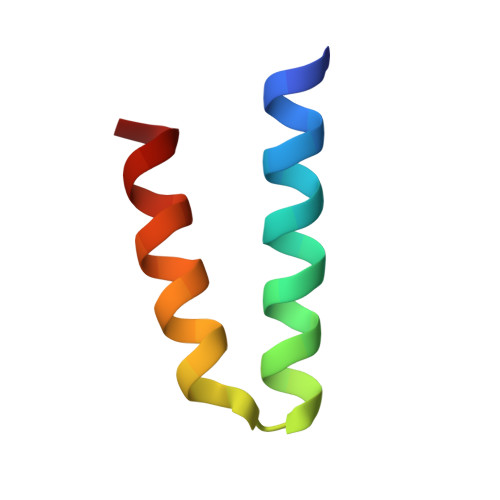
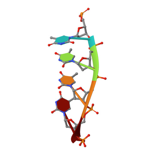
    UvrB is the damage recognition element of the highly conserved UvrABC pathway that functions in the removal of bulky DNA adducts. Pivotal to this is the formation of a damage detection complex that relies on the ability of UvrB to locate and sequester diverse lesions. Whilst structures of UvrB bound to DNA have recently been reported, none address the issue of lesion recognition. Here, we describe the crystal structure of UvrB bound to a pentanucleotide containing a single fluorescein-adducted thymine that reveals a unique mechanism for damage detection entirely dependent on the exclusion of lesions larger than an undamaged nucleotide.


  • Organizational Affiliation
    • The School of Crystallography and the Institute for Structural Molecular Biology, Birkbeck College, Malet Street, London WC1E 7HX, United Kingdom.

Macromolecule Content 

  • Total Structure Weight: 83.25 kDa 
  • Atom Count: 5,311 
  • Modeled Residue Count: 663 
  • Deposited Residue Count: 704 
  • Unique protein chains: 2
  • Unique nucleic acid chains: 1

Macromolecules


Find similar proteins by:|  3D Structure
Entity ID: 2
MoleculeChains  Sequence LengthOrganismDetailsImage
UvrABC system protein BB [auth A]661Bacillus subtilisMutation(s): 0 
Gene Names: uvrB
EC: 3.1
UniProt
Find proteins for P37954 (Bacillus subtilis (strain 168))
Explore P37954 
Go to UniProtKB:  P37954
Entity Groups
Sequence Clusters30% Identity50% Identity70% Identity90% Identity95% Identity100% Identity
UniProt GroupP37954
Sequence Annotations
Expand
Reference Sequence
Find similar proteins by:|  3D Structure
Entity ID: 3
MoleculeChains  Sequence LengthOrganismDetailsImage
UvrABC system protein BC [auth B]38Bacillus subtilisMutation(s): 0 
Gene Names: uvrB
EC: 3.1
UniProt
Find proteins for P37954 (Bacillus subtilis (strain 168))
Explore P37954 
Go to UniProtKB:  P37954
Entity Groups
Sequence Clusters30% Identity50% Identity70% Identity90% Identity95% Identity100% Identity
UniProt GroupP37954
Sequence Annotations
Expand
Reference Sequence
Find similar nucleic acids by:  Sequence
Entity ID: 1
MoleculeChains LengthOrganismImage
5'-D(P*TP*TP*TP*TP*T)-3'A [auth D]5N/A
Sequence Annotations
Expand
Reference Sequence

Small Molecules

Ligands 3 Unique
IDChains Name / Formula / InChI Key2D Diagram3D Interactions
ADP

Query on ADP


Download:Ideal Coordinates CCD File
F [auth A]ADENOSINE-5'-DIPHOSPHATE
C10 H15 N5 O10 P2
XTWYTFMLZFPYCI-KQYNXXCUSA-N
FLU

Query on FLU


Download:Ideal Coordinates CCD File
E [auth D]2-(6-HYDROXY-3-OXO-3H-XANTHEN-9-YL)-BENZOIC ACID
C20 H12 O5
YKGGGCXBWXHKIZ-UHFFFAOYSA-N
NML

Query on NML


Download:Ideal Coordinates CCD File
D
N-METHYLACETAMIDE
C3 H7 N O
OHLUUHNLEMFGTQ-UHFFFAOYSA-N

Experimental Data & Validation

Experimental Data

  • Method: X-RAY DIFFRACTION
  • Resolution: 2.95 Å
  • R-Value Free:  0.281 (Depositor) 
  • R-Value Work:  0.224 (Depositor) 
  • R-Value Observed: 0.227 (Depositor) 
Space Group: P 21 21 21
Unit Cell:
Length ( Å )Angle ( ˚ )
a = 74.41α = 90
b = 95.597β = 90
c = 97.878γ = 90
Software Package:
Software NamePurpose
REFMACrefinement
ADSCdata collection
MOSFLMdata reduction
CCP4data scaling
MOLREPphasing

Structure Validation

View Full Validation Report


Entry History 

Deposition Data

Revision History  (Full details and data files)

  • Version 1.0: 2007-01-16
    Type: Initial release
  • Version 1.1: 2008-05-01
    Changes: Version format compliance
  • Version 1.2: 2011-07-13
    Changes: Version format compliance
  • Version 1.3: 2023-10-25
    Changes: Data collection, Database references, Derived calculations, Refinement description