


Solution structure of the dithiolic glutaredoxin 2-C-Grx1 from the pathogen Trypanosoma brucei brucei
Sturlese, M., Lelli, M., Manta, B., Mammi, S., Comini, M.A., Bellanda, M.To be published.
Experimental Data Snapshot
wwPDB Validation 3D Report Full Report

Entity ID: 1 | |||||
|---|---|---|---|---|---|
| Molecule | Chains | Sequence Length | Organism | Details | Image |
| Dithiol glutaredoxin 1 | 97 | Trypanosoma brucei | Mutation(s): 0 Gene Names: grx1, TbgDal_XI1450 | 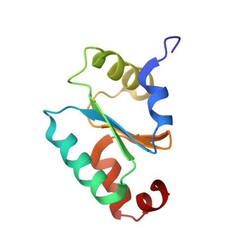 | |
UniProt | |||||
Entity Groups | |||||
| Sequence Clusters | 30% Identity50% Identity70% Identity90% Identity95% Identity100% Identity | ||||
| UniProt Group | D0A5S8 | ||||
Sequence AnnotationsExpand | |||||
Reference Sequence | |||||


