A Novel AT-Rich DNA Recognition Mechanism for Bacterial Xenogeneic Silencer MvaT.
Ding, P., McFarland, K.A., Jin, S., Tong, G., Duan, B., Yang, A., Hughes, T.R., Liu, J., Dove, S.L., Navarre, W.W., Xia, B.(2015) PLoS Pathog 11: e1004967-e1004967
- PubMed: 26068099 Search on PubMedSearch on PubMed Central
- DOI: https://doi.org/10.1371/journal.ppat.1004967
- Primary Citation Related Structures:
2MXE, 2MXF - PubMed Abstract:
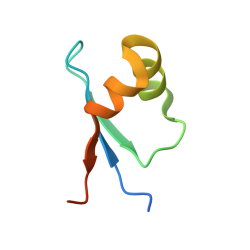
Bacterial xenogeneic silencing proteins selectively bind to and silence expression from many AT rich regions of the chromosome. They serve as master regulators of horizontally acquired DNA, including a large number of virulence genes. To date, three distinct families of xenogeneic silencers have been identified: H-NS of Proteobacteria, Lsr2 of the Actinomycetes, and MvaT of Pseudomonas sp. Although H-NS and Lsr2 family proteins are structurally different, they all recognize the AT-rich DNA minor groove through a common AT-hook-like motif, which is absent in the MvaT family. Thus, the DNA binding mechanism of MvaT has not been determined. Here, we report the characteristics of DNA sequences targeted by MvaT with protein binding microarrays, which indicates that MvaT prefers binding flexible DNA sequences with multiple TpA steps. We demonstrate that there are clear differences in sequence preferences between MvaT and the other two xenogeneic silencer families. We also determined the structure of the DNA-binding domain of MvaT in complex with a high affinity DNA dodecamer using solution NMR. This is the first experimental structure of a xenogeneic silencer in complex with DNA, which reveals that MvaT recognizes the AT-rich DNA both through base readout by an "AT-pincer" motif inserted into the minor groove and through shape readout by multiple lysine side chains interacting with the DNA sugar-phosphate backbone. Mutations of key MvaT residues for DNA binding confirm their importance with both in vitro and in vivo assays. This novel DNA binding mode enables MvaT to better tolerate GC-base pair interruptions in the binding site and less prefer A tract DNA when compared to H-NS and Lsr2. Comparison of MvaT with other bacterial xenogeneic silencers provides a clear picture that nature has evolved unique solutions for different bacterial genera to distinguish foreign from self DNA.
- Beijing Nuclear Magnetic Resonance Center, School of Life Sciences, College of Chemistry and Molecular Engineering, Peking University, Beijing, China.
Organizational Affiliation: