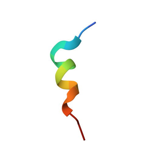
Monitoring Ligand-Induced Protein Ordering in Drug Discovery.
Grace, C.R., Ban, D., Min, J., Mayasundari, A., Min, L., Finch, K.E., Griffiths, L., Bharatham, N., Bashford, D., Kiplin Guy, R., Dyer, M.A., Kriwacki, R.W.(2016) J Mol Biology 428: 1290-1303
- PubMed: 26812210
- DOI: https://doi.org/10.1016/j.jmb.2016.01.016
- Primary Citation of Related Structures:
2MWY, 2N06, 2N0U, 2N0W, 2N14 - PubMed Abstract:
While the gene for p53 is mutated in many human cancers causing loss of function, many others maintain a wild-type gene but exhibit reduced p53 tumor suppressor activity through overexpression of the negative regulators, Mdm2 and/or MdmX. For the latter mechanism of loss of function, the activity of endogenous p53 can be restored through inhibition of Mdm2 or MdmX with small molecules. We previously reported a series of compounds based upon the Nutlin-3 chemical scaffold that bind to both MdmX and Mdm2 [Vara, B. A. et al. (2014) Organocatalytic, diastereo- and enantioselective synthesis of nonsymmetric cis-stilbene diamines: A platform for the preparation of single-enantiomer cis-imidazolines for protein-protein inhibition. J. Org. Chem. 79, 6913-6938]. Here we present the first solution structures based on data from NMR spectroscopy for MdmX in complex with four of these compounds and compare them with the MdmX:p53 complex. A p53-derived peptide binds with high affinity (Kd value of 150nM) and causes the formation of an extensive network of hydrogen bonds within MdmX; this constitutes the induction of order within MdmX through ligand binding. In contrast, the compounds bind more weakly (Kd values from 600nM to 12μM) and induce an incomplete hydrogen bond network within MdmX. Despite relatively weak binding, the four compounds activated p53 and induced p21(Cip1) expression in retinoblastoma cell lines that overexpress MdmX, suggesting that they specifically target MdmX and/or Mdm2. Our results document structure-activity relationships for lead-like small molecules targeting MdmX and suggest a strategy for their further optimization in the future by using NMR spectroscopy to monitor small-molecule-induced protein order as manifested through hydrogen bond formation.
- Department of Structural Biology, St. Jude Children's Research Hospital, 262 Danny Thomas Place, Memphis, TN 38105, USA.
Organizational Affiliation:


