2MNU | pdb_00002mnu

Backbone and side chain 1H, 13C, and 15N Chemical Shift Assignments for EDB and specific binding aptide


Experimental Data Snapshot

  • Method: SOLUTION NMR
  • Conformers Calculated: 50 
  • Conformers Submitted: 20 
  • Selection Criteria: structures with the least restraint violations 

wwPDB Validation 3D Report Full Report

Validation slider image for 2MNU

This is version 1.2 of the entry. See complete history

Literature

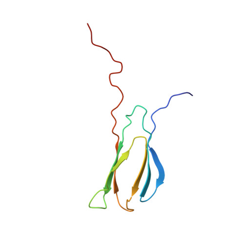
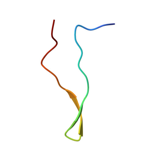
An Unusual Protein-Protein Interaction through Coupled Unfolding and Binding

Yu, T.K.Shin, S.A.Kim, E.H.Kim, S.Ryu, K.S.Cheong, H.Ahn, H.C.Jon, S.Suh, J.Y.

(2014) Angew Chem Int Ed Engl 53: 9784-9787

  • DOI: https://doi.org/10.1002/anie.201404750
  • Primary Citation Related Structures: 
    2MNU

  • PubMed Abstract: 

    Aptides, a novel class of high-affinity peptides, recognize diverse molecular targets with high affinity and specificity. The solution structure of the aptide APT specifically bound to fibronectin extradomain B (EDB), which represents an unusual protein-protein interaction that involves coupled unfolding and binding, is reported. APT binding is accompanied by unfolding of the C-terminal β strand of EDB, thereby permitting APT to interact with the freshly exposed hydrophobic interior surfaces of EDB. The β-hairpin scaffold of APT drives the interaction by a β-strand displacement mechanism, such that an intramolecular β sheet is replaced by an intermolecular β sheet. The unfolding of EDB perturbs the tight domain association between EDB and FN8 of fibronectin, thus highlighting its potential use as a scaffold that switches between stretched and bent conformations.


  • Organizational Affiliation
    • Biomodulation Major, Department of Agricultural Biotechnology, Seoul National University, 1 Gwanak-ro, Gwanak-gu, Seoul 151-921 (South Korea).

Macromolecule Content 

  • Total Structure Weight: 12.94 kDa 
  • Atom Count: 913 
  • Modeled Residue Count: 119 
  • Deposited Residue Count: 119 
  • Unique protein chains: 2

Macromolecules

Find similar proteins by:|  3D Structure
Entity ID: 1
MoleculeChains  Sequence LengthOrganismDetailsImage
EDB93Homo sapiensMutation(s): 0 
UniProt & NIH Common Fund Data Resources
Find proteins for P02751 (Homo sapiens)
Explore P02751 
Go to UniProtKB:  P02751
PHAROS:  P02751
GTEx:  ENSG00000115414 
Entity Groups
Sequence Clusters30% Identity50% Identity70% Identity90% Identity95% Identity100% Identity
UniProt GroupP02751
Sequence Annotations
Expand
Reference Sequence
Find similar proteins by:|  3D Structure
Entity ID: 2
MoleculeChains  Sequence LengthOrganismDetailsImage
APT26Homo sapiensMutation(s): 0 

Experimental Data & Validation

Experimental Data

  • Method: SOLUTION NMR
  • Conformers Calculated: 50 
  • Conformers Submitted: 20 
  • Selection Criteria: structures with the least restraint violations 

Structure Validation

View Full Validation Report


Entry History 

Deposition Data

  • Released Date: 2014-09-24 
  • Deposition Author(s): Suh, J., Yu, T.

Revision History  (Full details and data files)

  • Version 1.0: 2014-09-24
    Type: Initial release
  • Version 1.1: 2023-06-14
    Changes: Data collection, Database references, Other
  • Version 1.2: 2024-05-15
    Changes: Data collection, Database references