Comparative Sequence, Structure and Redox Analyses of Klebsiella pneumoniae DsbA Show That Anti-Virulence Target DsbA Enzymes Fall into Distinct Classes.
Kurth, F., Rimmer, K., Premkumar, L., Mohanty, B., Duprez, W., Halili, M.A., Shouldice, S.R., Heras, B., Fairlie, D.P., Scanlon, M.J., Martin, J.L.(2013) PLoS One 8: e80210-e80210
- PubMed: 24244651 Search on PubMedSearch on PubMed Central
- DOI: https://doi.org/10.1371/journal.pone.0080210
- Primary Citation Related Structures:
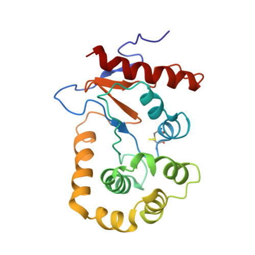
2MBS, 4MCU - PubMed Abstract:
Bacterial DsbA enzymes catalyze oxidative folding of virulence factors, and have been identified as targets for antivirulence drugs. However, DsbA enzymes characterized to date exhibit a wide spectrum of redox properties and divergent structural features compared to the prototypical DsbA enzyme of Escherichia coli DsbA (EcDsbA). Nonetheless, sequence analysis shows that DsbAs are more highly conserved than their known substrate virulence factors, highlighting the potential to inhibit virulence across a range of organisms by targeting DsbA. For example, Salmonella enterica typhimurium (SeDsbA, 86 % sequence identity to EcDsbA) shares almost identical structural, surface and redox properties. Using comparative sequence and structure analysis we predicted that five other bacterial DsbAs would share these properties. To confirm this, we characterized Klebsiella pneumoniae DsbA (KpDsbA, 81 % identity to EcDsbA). As expected, the redox properties, structure and surface features (from crystal and NMR data) of KpDsbA were almost identical to those of EcDsbA and SeDsbA. Moreover, KpDsbA and EcDsbA bind peptides derived from their respective DsbBs with almost equal affinity, supporting the notion that compounds designed to inhibit EcDsbA will also inhibit KpDsbA. Taken together, our data show that DsbAs fall into different classes; that DsbAs within a class may be predicted by sequence analysis of binding loops; that DsbAs within a class are able to complement one another in vivo and that compounds designed to inhibit EcDsbA are likely to inhibit DsbAs within the same class.
- Division of Chemistry and Structural Biology, Institute for Molecular Bioscience, The University of Queensland, Brisbane, Queensland, Australia.
Organizational Affiliation: