Solution structure of a mutant of the triheme cytochrome PpcA from Geobacter sulfurreducens sheds light on the role of the conserved aromatic residue F15.
Dantas, J.M., Morgado, L., Pokkuluri, P.R., Turner, D.L., Salgueiro, C.A.(2013) Biochim Biophys Acta 1827: 484-492
- PubMed: 23313804 Search on PubMed
- DOI: https://doi.org/10.1016/j.bbabio.2012.12.008
- Primary Citation Related Structures:
2LZZ - PubMed Abstract:
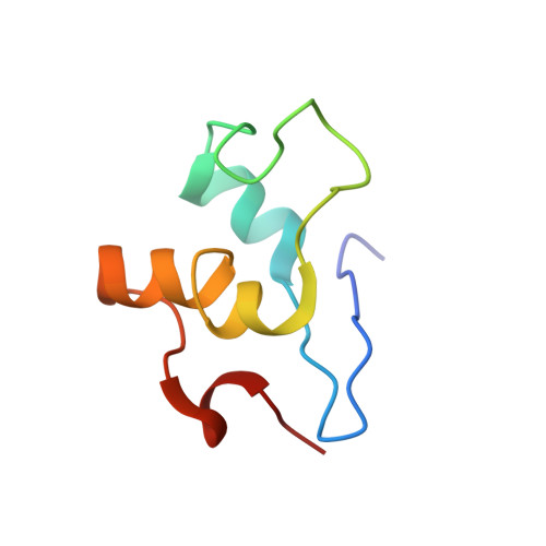
Extracellular electron transfer is one of the physiological hallmarks of Geobacteraceae. Most of the Geobacter species encode for more than 100 c-type cytochromes which are, in general, poorly conserved between individual species. An exception to this is the PpcA family of periplasmic triheme c-type cytochromes, which are the most abundant proteins in these bacteria. The functional characterization of PpcA showed that it has the necessary properties to couple electron/proton transfer, a fundamental step for ATP synthesis. The detailed thermodynamic characterization of a PpcA mutant, in which the strictly conserved residue phenylalanine 15 was replaced by leucine, showed that the global redox network of cooperativities among heme groups is altered, preventing the mutant from performing a concerted electron/proton transfer. In this work, we determined the solution structure of PpcA F15L mutant in the fully reduced state using NMR spectroscopy by producing (15)N-labeled protein. In addition, pH-dependent conformational changes were mapped onto the structure. The mutant structure obtained is well defined, with an average pairwise root-mean-square deviation of 0.36Å for the backbone atoms and 1.14Å for all heavy atoms. Comparison between the mutant and wild-type structures elucidated the contribution of phenylalanine 15 in the modulation of the functional properties of PpcA.
- Requimte-CQFB, Departamento de Química, Faculdade de Ciências e Tecnologia, Universidade Nova de Lisboa, Campus Caparica, 2829-516 Caparica, Portugal.
Organizational Affiliation: