Download Mendeley
Lanthanide binding and IgG affinity construct: Potential applications in solution NMR, MRI, and luminescence microscopy.
Barb, A.W., Ho, T.G., Flanagan-Steet, H., Prestegard, J.H.(2012) Protein Sci 21: 1456-1466
- PubMed: 22851279 Search on PubMedSearch on PubMed Central
- DOI: https://doi.org/10.1002/pro.2133
- Primary Citation Related Structures:
2LR2 - PubMed Abstract:
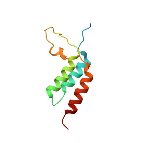
Paramagnetic lanthanide ions when bound to proteins offer great potential for structural investigations that utilize solution nuclear magnetic resonance spectroscopy, magnetic resonance imaging, or optical microscopy. However, many proteins do not have native metal ion binding sites and engineering a chimeric protein to bind an ion while retaining affinity for a protein of interest represents a significant challenge. Here we report the characterization of an immunoglobulin G-binding protein redesigned to include a lanthanide binding motif in place of a loop between two helices (Z-L2LBT). It was shown to bind Tb³⁺ with 130 nM affinity. Ions such as Dy³⁺, Yb³⁺, and Ce³⁺ produce paramagnetic effects on NMR spectra and the utility of these effects is illustrated by their use in determining a structural model of the metal-complexed Z-L2LBT protein and a preliminary characterization of the dynamic distribution of IgG Fc glycan positions. Furthermore, this designed protein is demonstrated to be a novel IgG-binding reagent for magnetic resonance imaging (Z-L2LBT:Gd³⁺ complex) and luminescence microscopy (Z-L2LBT: Tb³⁺ complex).
- Complex Carbohydrate Research Center, The University of Georgia, Athens, Georgia, USA.
Organizational Affiliation: