Structure, dynamics and domain organization of the repeat protein Cin1 from the apple scab fungus.
Mesarich, C.H., Schmitz, M., Tremouilhac, P., McGillivray, D.J., Templeton, M.D., Dingley, A.J.(2012) Biochim Biophys Acta 1824: 1118-1128
- PubMed: 22771296 Search on PubMed
- DOI: https://doi.org/10.1016/j.bbapap.2012.06.015
- Primary Citation Related Structures:
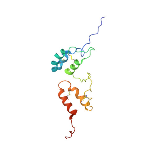
2LHT - PubMed Abstract:
Venturia inaequalis is a hemi-biotrophic fungus that causes scab disease of apple. A recently-identified gene from this fungus, cin1 (cellophane-induced 1), is up-regulated over 1000-fold in planta and considerably on cellophane membranes, and encodes a cysteine-rich secreted protein of 523 residues with eight imperfect tandem repeats of ~60 amino acids. The Cin1 sequence has no homology to known proteins and appears to be genus-specific; however, Cin1 repeats and other repeat domains may be structurally similar. An NMR-derived structure of the first two repeat domains of Cin1 (Cin1-D1D2) and a low-resolution model of the full-length protein (Cin1-FL) using SAXS data were determined. The structure of Cin1-D1D2 reveals that each domain comprises a core helix-loop-helix (HLH) motif as part of a three-helix bundle, and is stabilized by two intra-domain disulfide bonds. Cin1-D1D2 adopts a unique protein fold as DALI and PDBeFOLD analysis identified no structural homology. A (15)N backbone NMR dynamic analysis of Cin1-D1D2 showed that a short stretch of the inter-domain linker has large amplitude motions that give rise to reciprocal domain-domain mobility. This observation was supported by SAXS data modeling, where the scattering length density envelope remains thick at the domain-domain boundary, indicative of inter-domain dynamics. Cin1-FL SAXS data models a loosely-packed arrangement of domains, rather than the canonical parallel packing of adjacent HLH repeats observed in α-solenoid repeat proteins. Together, these data suggest that the repeat domains of Cin1 display a "beads-on-a-string" organization with inherent inter-domain flexibility that is likely to facilitate interactions with target ligands.
- School of Biological Sciences, The University of Auckland, Private Bag 92 019, Auckland 1142, New Zealand.
Organizational Affiliation: