Structure and function of the complete internal fusion loop from Ebolavirus glycoprotein 2.
Gregory, S.M., Harada, E., Liang, B., Delos, S.E., White, J.M., Tamm, L.K.(2011) Proc Natl Acad Sci U S A 108: 11211-11216
- PubMed: 21690393 Search on PubMedSearch on PubMed Central
- DOI: https://doi.org/10.1073/pnas.1104760108
- Primary Citation Related Structures:
2LCY, 2LCZ - PubMed Abstract:
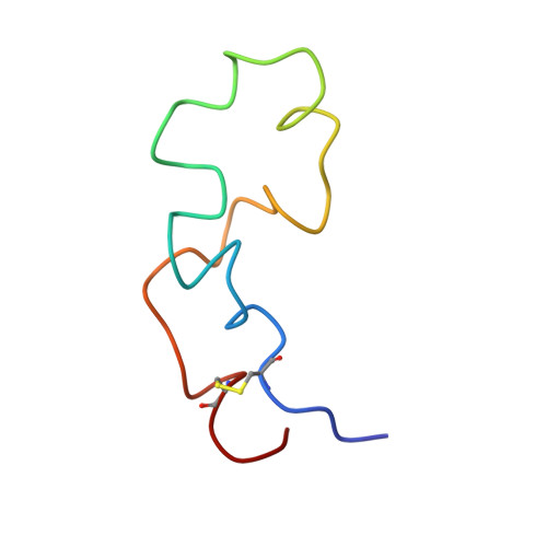
Ebolavirus (Ebov), an enveloped virus of the family Filoviridae, causes hemorrhagic fever in humans and nonhuman primates. The viral glycoprotein (GP) is solely responsible for virus-host membrane fusion, but how it does so remains elusive. Fusion occurs after virions reach an endosomal compartment where GP is proteolytically primed by cathepsins. Fusion by primed GP is governed by an internal fusion loop found in GP2, the fusion subunit. This fusion loop contains a stretch of hydrophobic residues, some of which have been shown to be critical for GP-mediated infection. Here we present liposome fusion data and NMR structures for a complete (54-residue) disulfide-bonded internal fusion loop (Ebov FL) in a membrane mimetic. The Ebov FL induced rapid fusion of liposomes of varying compositions at pH values at or below 5.5. Consistently, circular dichroism experiments indicated that the α-helical content of the Ebov FL in the presence of either lipid-mimetic micelles or small liposomes increases in samples exposed to pH ≤5.5. NMR structures in dodecylphosphocholine micelles at pH 7.0 and 5.5 revealed a conformational change from a relatively flat extended loop structure at pH 7.0 to a structure with an ∼90° bend at pH 5.5. Induction of the bend at low pH reorients and compacts the hydrophobic patch at the tip of the FL. We propose that these changes facilitate disruption of lipids at the site of virus-host cell membrane contact and, hence, initiate Ebov fusion.
- Center for Membrane Biology, University of Virginia, Charlottesville, VA 22908, USA.
Organizational Affiliation: