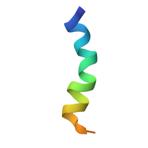
Shallow boomerang-shaped influenza hemagglutinin G13A mutant structure promotes leaky membrane fusion.
Lai, A.L., Tamm, L.K.(2010) J Biological Chem 285: 37467-37475
- PubMed: 20826788 Search on PubMedSearch on PubMed Central
- DOI: https://doi.org/10.1074/jbc.M110.153700
- Primary Citation Related Structures:
2L4G - PubMed Abstract:
Our previous studies showed that an angled boomerang-shaped structure of the influenza hemagglutinin (HA) fusion domain is critical for virus entry into host cells by membrane fusion. Because the acute angle of ∼105° of the wild-type fusion domain promotes efficient non-leaky membrane fusion, we asked whether different angles would still support fusion and thus facilitate virus entry. Here, we show that the G13A fusion domain mutant produces a new leaky fusion phenotype. The mutant fusion domain structure was solved by NMR spectroscopy in a lipid environment at fusion pH. The mutant adopted a boomerang structure similar to that of wild type but with a shallower kink angle of ∼150°. G13A perturbed the structure of model membranes to a lesser degree than wild type but to a greater degree than non-fusogenic fusion domain mutants. The strength of G13A binding to lipid bilayers was also intermediate between that of wild type and non-fusogenic mutants. These membrane interactions provide a clear link between structure and function of influenza fusion domains: an acute angle is required to promote clean non-leaky fusion suitable for virus entry presumably by interaction of the fusion domain with the transmembrane domain deep in the lipid bilayer. A shallower angle perturbs the bilayer of the target membrane so that it becomes leaky and unable to form a clean fusion pore. Mutants with no fixed boomerang angle interacted with bilayers weakly and did not promote any fusion or membrane perturbation.
- Center for Membrane Biology, Department of Molecular Physiology and Biological Physics, University of Virginia, Charlottesville, Virginia 22903-0886, USA.
Organizational Affiliation: