2KYT | pdb_00002kyt

Solution structure of the H-REV107 N-terminal domain


Experimental Data Snapshot

  • Method: SOLUTION NMR
  • Conformers Calculated: 100 
  • Conformers Submitted: 20 
  • Selection Criteria: structures with the lowest energy 

wwPDB Validation 3D Report Full Report

Validation slider image for 2KYT

This is version 1.3 of the entry. See complete history

Literature

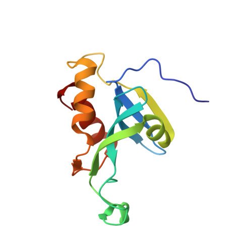
Solution structure of the N-terminal catalytic domain of human H-REV107--a novel circular permutated NlpC/P60 domain

Ren, X.Lin, J.Jin, C.Xia, B.

(2010) FEBS Lett 584: 4222-4226

  • DOI: https://doi.org/10.1016/j.febslet.2010.09.015
  • Primary Citation Related Structures: 
    2KYT

  • PubMed Abstract: 

    H-REV107 is a Ca(2+)-independent phospholipase A(1/2), and it is also a pro-apoptosis protein belonging to the novel class II tumor suppressor family, H-REV107-like family. Here we report the solution structure of the N-terminal catalytic domain of human H-REV107, which has a similar architecture to classical NlpC/P60 domains, even though their fold topologies are different due to circular permutation in the primary sequence. The phospholipase active site possesses a structurally conserved Cys-His-His catalytic triad as found in NlpC/P60 peptidases, indicating H-REV107 should adopt a similar catalytic mechanism towards phospholipid substrates to that of NlpC/P60 peptidases towards peptides. As H-REV107 is highly similar to lecithin retinol acyltransferase, our study also provides structural insight to this essential enzyme in retinol metabolism.


  • Organizational Affiliation
    • Beijing Nuclear Magnetic Resonance Center, Peking University, Beijing, China.

Macromolecule Content 

  • Total Structure Weight: 13.97 kDa 
  • Atom Count: 984 
  • Modeled Residue Count: 125 
  • Deposited Residue Count: 125 
  • Unique protein chains: 1

Macromolecules

Find similar proteins by:|  3D Structure
Entity ID: 1
MoleculeChains  Sequence LengthOrganismDetailsImage
Group XVI phospholipase A2125Homo sapiensMutation(s): 0 
Gene Names: PLA2G16HRASLS3HREV107
EC: 3.1.1.4 (PDB Primary Data), 2.3.1 (UniProt), 3.1.1.32 (UniProt)
Membrane Entity: Yes 
UniProt & NIH Common Fund Data Resources
Find proteins for P53816 (Homo sapiens)
Explore P53816 
Go to UniProtKB:  P53816
PHAROS:  P53816
GTEx:  ENSG00000176485 
Entity Groups
Sequence Clusters30% Identity50% Identity70% Identity90% Identity95% Identity100% Identity
UniProt GroupP53816
Sequence Annotations
Expand
Reference Sequence

Experimental Data & Validation

Experimental Data

  • Method: SOLUTION NMR
  • Conformers Calculated: 100 
  • Conformers Submitted: 20 
  • Selection Criteria: structures with the lowest energy 

Structure Validation

View Full Validation Report


Entry History 

Deposition Data

Revision History  (Full details and data files)

  • Version 1.0: 2010-11-03
    Type: Initial release
  • Version 1.1: 2011-07-13
    Changes: Version format compliance
  • Version 1.2: 2016-12-21
    Changes: Structure summary
  • Version 1.3: 2024-05-01
    Changes: Data collection, Database references