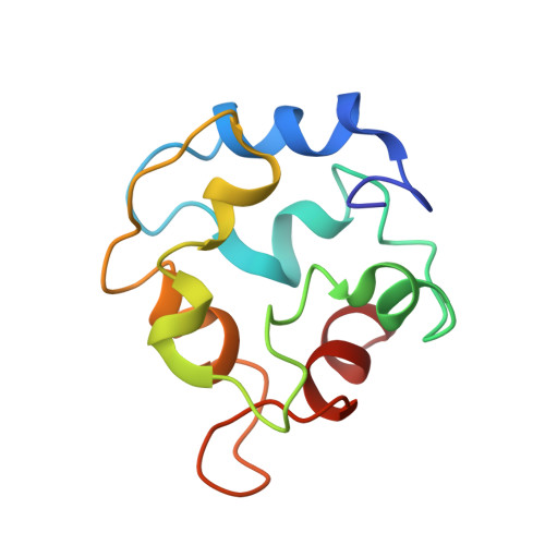
Solution structures of chicken parvalbumin 3 in the Ca(2+) -free and Ca(2+) -bound states.
Henzl, M.T., Tanner, J.J., Tan, A.(2011) Proteins 79: 752-764
- PubMed: 21287610 Search on PubMed
- DOI: https://doi.org/10.1002/prot.22915
- Primary Citation Related Structures:
2KYC, 2KYF - PubMed Abstract:
Birds express two β-parvalbumin isoforms, parvalbumin 3 and avian thymic hormone (ATH). Parvalbumin 3 from chicken (CPV3) is identical to rat β-parvalbumin (β-PV) at 75 of 108 residues. CPV3 displays intermediate Ca(2+) affinity--higher than that of rat β-parvalbumin, but lower than that of ATH. As in rat β-PV, the attenuation of affinity is associated primarily with the CD site (residues 41-70), rather than the EF site (residues 80-108). Structural data for rat α- and β-parvalbumins suggest that divalent ion affinity is correlated with the similarity of the unliganded and Ca(2+)-bound conformations. We herein present a comparison of the solution structures of Ca(2+)-free and Ca(2+)-bound CPV3. Although the structures are generally similar, the conformations of residues 47 to 50 differ markedly in the two protein forms. These residues are located in the C helix, proximal to the CD binding loop. In response to Ca(2+) removal, F47 experiences much greater solvent accessibility. The side-chain of R48 assumes a position between the C and D helices, adjacent to R69. Significantly, I49 adopts an interior position in the unliganded protein that allows association with the side-chain of L50. Concomitantly, the realignment of F66 and F70 facilitates their interaction with I49 and reduces their contact with residues in the N-terminal AB domain. This reorganization of the hydrophobic core, although less profound, is nevertheless reminiscent of that observed in rat β-PV. The results lend further support to the idea that Ca(2+) affinity correlates with the structural similarity of the apo- and bound parvalbumin conformations.
- Department of Biochemistry, University of Missouri, Columbia, Missouri 65211, USA. henzlm@missouri.edu
Organizational Affiliation: