2KX7 | pdb_00002kx7

Solution structure of the E.coli RcsD-ABL domain (residues 688-795)


Experimental Data Snapshot

  • Method: SOLUTION NMR
  • Conformers Calculated: 50 
  • Conformers Submitted: 25 
  • Selection Criteria: structures with the lowest energy 

wwPDB Validation 3D Report Full Report

Validation slider image for 2KX7

This is version 1.2 of the entry. See complete history

Literature

Structural Insights into Rcs Phosphotransfer: The Newly Identified RcsD-ABL Domain Enhances Interaction with the Response Regulator RcsB.

Schmoe, K.Rogov, V.V.Rogova, N.Y.Lohr, F.Guntert, P.Bernhard, F.Dotsch, V.

(2011) Structure 19: 577-587

  • DOI: https://doi.org/10.1016/j.str.2011.01.012
  • Primary Citation Related Structures: 
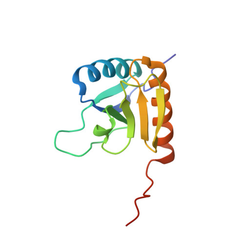
    2KX7

  • PubMed Abstract: 

    The Rcs-signaling system is one of the most remarkable phosphorelay pathways in Enterobacteriaceae, comprising several membrane-bound and soluble proteins. Within the complex phosphotransfer pathway, the histidine phosphotransferase (HPt) domain of the RcsD membrane-bound component serves as a crucial factor in modulating the phosphorylation state of the transcription factor RcsB. We have identified a new domain, RcsD-ABL, located N terminally to RcsD-HPt that interacts with RcsB as well. We have determined its structure, characterized its interaction interface with RcsB, and built a structural model of the complex of the RcsD-ABL domain with RcsB. Our results indicate that the effector domain of RcsB, which normally binds to DNA, is recognized by RcsD-ABL, whereas the HPt domain interacts with the phosphoreceiver domain of RcsB.


  • Organizational Affiliation
    • Institute of Biophysical Chemistry and Center for Biomolecular Magnetic Resonance, Goethe University, Max-von-Laue-Strasse 9, 60438 Frankfurt/Main, Germany.

Macromolecule Content 

  • Total Structure Weight: 13.17 kDa 
  • Atom Count: 858 
  • Modeled Residue Count: 111 
  • Deposited Residue Count: 117 
  • Unique protein chains: 1

Macromolecules

Find similar proteins by:|  3D Structure
Entity ID: 1
MoleculeChains  Sequence LengthOrganismDetailsImage
Sensor-like histidine kinase yojN117Escherichia coli K-12Mutation(s): 0 
Gene Names: b2216JW2204yojNyojPyojQ
EC: 2.7.13.3 (PDB Primary Data), 2.7.2 (UniProt)
UniProt
Find proteins for P39838 (Escherichia coli (strain K12))
Explore P39838 
Go to UniProtKB:  P39838
Entity Groups
Sequence Clusters30% Identity50% Identity70% Identity90% Identity95% Identity100% Identity
UniProt GroupP39838
Sequence Annotations
Expand
Reference Sequence

Experimental Data & Validation

Experimental Data

  • Method: SOLUTION NMR
  • Conformers Calculated: 50 
  • Conformers Submitted: 25 
  • Selection Criteria: structures with the lowest energy 

Structure Validation

View Full Validation Report


Entry History 

Deposition Data

Revision History  (Full details and data files)

  • Version 1.0: 2011-04-06
    Type: Initial release
  • Version 1.1: 2011-07-13
    Changes: Version format compliance
  • Version 1.2: 2024-05-01
    Changes: Data collection, Database references