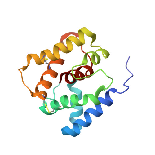
NMR Structure of Navel Orangeworm Moth Pheromone-Binding Protein (AtraPBP1): Implications for pH-Sensitive Pheromone Detection .
Xu, X., Xu, W., Rayo, J., Ishida, Y., Leal, W.S., Ames, J.B.(2010) Biochemistry 49: 1469-1476
- PubMed: 20088570
- DOI: https://doi.org/10.1021/bi9020132
- Primary Citation of Related Structures:
2KPH - PubMed Abstract:
The navel orangeworm, Amyelois transitella (Walker), is an agricultural insect pest that can be controlled by disrupting male-female communication with sex pheromones, a technique known as mating disruption. Insect pheromone-binding proteins (PBPs) provide fast transport of hydrophobic pheromones through the aqueous sensillar lymph and promote sensitive delivery of pheromones to receptors. Here we present the three-dimensional structure of a PBP from A. transitella (AtraPBP1) in solution at pH 4.5 determined by nuclear magnetic resonance (NMR) spectroscopy. Pulsed-field gradient NMR diffusion experiments, multiangle light scattering, and (15)N NMR relaxation analysis indicate that AtraPBP1 forms a stable monomer in solution at pH 4.5 in contrast to forming mostly dimers at pH 7. The NMR structure of AtraPBP1 at pH 4.5 contains seven alpha-helices (alpha1, L8-L23; alpha2, D27-F36; alpha3, R46-V62; alpha4, A73-M78; alpha5, D84-S100; alpha6, R107-L125; alpha7, M131-E141) that adopt an overall main-chain fold similar to that of PBPs found in Antheraea polyphemus and Bombyx mori. The AtraPBP1 structure is stabilized by three disulfide bonds formed by C19/C54, C50/C108, and C97/C117 and salt bridges formed by H69/E60, H70/E57, H80/E132, H95/E141, and H123/D40. All five His residues are cationic at pH 4.5, whereas H80 and H95 become neutral at pH 7.0. The C-terminal helix (alpha7) contains hydrophobic residues (M131, V133, V134, V135, V138, L139, and A140) that contact conserved residues (W37, L59, A73, F76, A77, I94, V111, and V115) suggested to interact with bound pheromone. Our NMR studies reveal that acid-induced formation of the C-terminal helix at pH 4.5 is triggered by a histidine protonation switch that promotes rapid release of bound pheromone under acidic conditions.
- Department of Chemistry, University of California, Davis, California 95616, USA.
Organizational Affiliation:


