A minimal sequence code for switching protein structure and function.
Alexander, P.A., He, Y., Chen, Y., Orban, J., Bryan, P.N.(2009) Proc Natl Acad Sci U S A 106: 21149-21154
- PubMed: 19923431 Search on PubMedSearch on PubMed Central
- DOI: https://doi.org/10.1073/pnas.0906408106
- Primary Citation Related Structures:
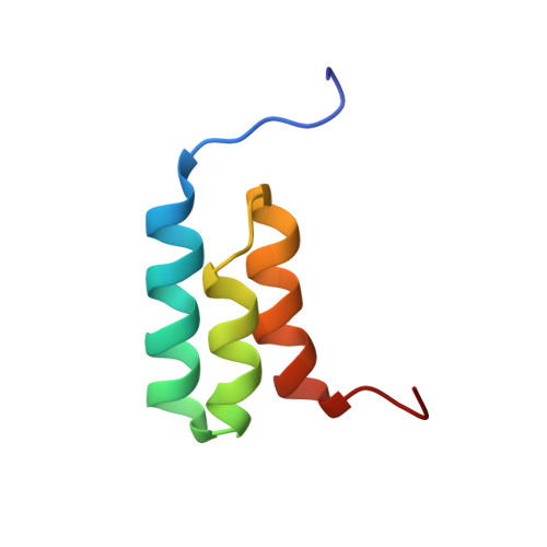
2KDL, 2KDM - PubMed Abstract:
We present here a structural and mechanistic description of how a protein changes its fold and function, mutation by mutation. Our approach was to create 2 proteins that (i) are stably folded into 2 different folds, (ii) have 2 different functions, and (iii) are very similar in sequence. In this simplified sequence space we explore the mutational path from one fold to another. We show that an IgG-binding, 4beta+alpha fold can be transformed into an albumin-binding, 3-alpha fold via a mutational pathway in which neither function nor native structure is completely lost. The stabilities of all mutants along the pathway are evaluated, key high-resolution structures are determined by NMR, and an explanation of the switching mechanism is provided. We show that the conformational switch from 4beta+alpha to 3-alpha structure can occur via a single amino acid substitution. On one side of the switch point, the 4beta+alpha fold is >90% populated (pH 7.2, 20 degrees C). A single mutation switches the conformation to the 3-alpha fold, which is >90% populated (pH 7.2, 20 degrees C). We further show that a bifunctional protein exists at the switch point with affinity for both IgG and albumin.
- Center for Advanced Research in Biotechnology, University of Maryland Biotechnology Institute, Rockville, MD 20850, USA.
Organizational Affiliation: