2K7N | pdb_00002k7n

Solution structure of the PPIL1 bound to a fragment of SKIP


Experimental Data Snapshot

  • Method: SOLUTION NMR
  • Conformers Calculated: 400 
  • Conformers Submitted: 20 
  • Selection Criteria: 20 structures for lowest energy 

wwPDB Validation 3D Report Full Report

Validation slider image for 2K7N

This is version 1.4 of the entry. See complete history

Literature

Solution Structure of PPIL1 Bound to the Fragment of SKIP Shown Disorder-Order Transition Induced by Protein Binding

Wang, X.Wu, J.Shi, Y.

To be published.

Macromolecule Content 

  • Total Structure Weight: 21.53 kDa 
  • Atom Count: 1,514 
  • Modeled Residue Count: 203 
  • Deposited Residue Count: 203 
  • Unique protein chains: 1

Macromolecules

Find similar proteins by:|  3D Structure
Entity ID: 1
MoleculeChains  Sequence LengthOrganismDetailsImage
Peptidyl-prolyl cis-trans isomerase-like 1203Homo sapiensMutation(s): 0 
Gene Names: PPIL1CYPL1CGI-124UNQ2425/PRO4984
EC: 5.2.1.8
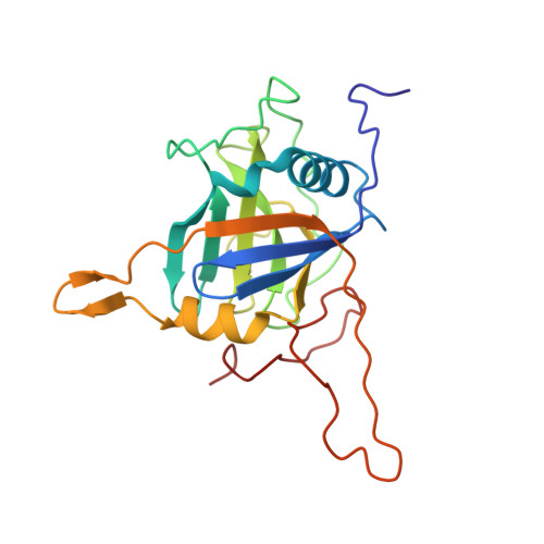
UniProt & NIH Common Fund Data Resources
Find proteins for Q9Y3C6 (Homo sapiens)
Explore Q9Y3C6 
Go to UniProtKB:  Q9Y3C6
PHAROS:  Q9Y3C6
GTEx:  ENSG00000137168 
Entity Groups
Sequence Clusters30% Identity50% Identity70% Identity90% Identity95% Identity100% Identity
UniProt GroupQ9Y3C6
Sequence Annotations
Expand
Reference Sequence

Experimental Data & Validation

Experimental Data

  • Method: SOLUTION NMR
  • Conformers Calculated: 400 
  • Conformers Submitted: 20 
  • Selection Criteria: 20 structures for lowest energy 

Structure Validation

View Full Validation Report


Entry History 

Deposition Data

Revision History  (Full details and data files)

  • Version 1.0: 2009-09-15
    Type: Initial release
  • Version 1.1: 2011-07-13
    Changes: Version format compliance
  • Version 1.2: 2020-02-26
    Changes: Database references, Derived calculations, Other
  • Version 1.3: 2023-06-14
    Changes: Database references, Other
  • Version 1.4: 2024-05-15
    Changes: Data collection, Database references