Solution structure of an active mutant of maize ribosome-inactivating protein (MOD) and its interaction with the ribosomal stalk protein P2.
Yang, Y., Mak, A.N., Shaw, P.C., Sze, K.H.(2010) J Mol Biology 395: 897-907
- PubMed: 19900464 Search on PubMed
- DOI: https://doi.org/10.1016/j.jmb.2009.10.051
- Primary Citation Related Structures:
2K6H - PubMed Abstract:
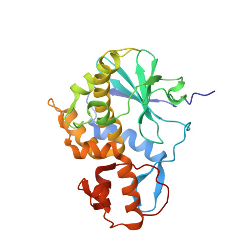
Ribosome-inactivating proteins (RIPs) are N-glycosidases that depurinate a specific adenine residue in the conserved sarcin/ricin loop of ribosomal RNA. This modification renders the ribosome unable to bind the elongation factors, thereby inhibiting the protein synthesis. Maize RIP, a type III RIP, is unique compared to the other type I and type II RIPs because it is synthesized as a precursor with a 25-residue internal inactivation region, which is removed in order to activate the protein. In this study, we describe the first solution structure of this type of RIP, a 28-kDa active mutant of maize RIP (MOD). The overall protein structure of MOD is comparable to those of the other type I RIPs and the A-chain of type II RIPs but shows significant differences in specific regions, including (1) shorter beta6 and alphaB segments, probably for accommodating easier substrate binding, and (2) an alpha-helix instead of an antiparallel beta-sheet in the C-terminal domain, which has been reported to be involved in binding ribosomal protein P2 in some RIPs. Furthermore, NMR chemical shift perturbation experiments revealed that the P2 binding site on MOD is located at the N-terminal domain near the internal inactivation region. This relocation of the P2 binding site can be rationalized by concerted changes in the electrostatic surface potential and 3D structures on the MOD protein and provides vital clues about the underlying molecular mechanism of this unique type of RIP.
- Chemistry Department and Open Laboratory of Chemical Biology, Institute of Molecular Technology for Drug Discovery and Synthesis, The University of Hong Kong, Pokfulam Road, Hong Kong, PR China.
Organizational Affiliation: