Structures of invisible, excited protein states by relaxation dispersion NMR spectroscopy
Vallurupalli, P., Hansen, D.F., Kay, L.E.(2008) Proc Natl Acad Sci U S A 105: 11766-11771
- PubMed: 18701719 Search on PubMedSearch on PubMed Central
- DOI: https://doi.org/10.1073/pnas.0804221105
- Primary Citation Related Structures:
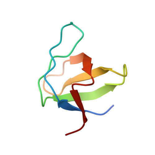
2K3B - PubMed Abstract:
Molecular function is often predicated on excursions between ground states and higher energy conformers that can play important roles in ligand binding, molecular recognition, enzyme catalysis, and protein folding. The tools of structural biology enable a detailed characterization of ground state structure and dynamics; however, studies of excited state conformations are more difficult because they are of low population and may exist only transiently. Here we describe an approach based on relaxation dispersion NMR spectroscopy in which structures of invisible, excited states are obtained from chemical shifts and residual anisotropic magnetic interactions. To establish the utility of the approach, we studied an exchanging protein (Abp1p SH3 domain)-ligand (Ark1p peptide) system, in which the peptide is added in only small amounts so that the ligand-bound form is invisible. From a collection of (15)N, (1)HN, (13)C(alpha), and (13)CO chemical shifts, along with (1)HN-(15)N, (1)H(alpha)-(13)C(alpha), and (1)HN-(13)CO residual dipolar couplings and (13)CO residual chemical shift anisotropies, all pertaining to the invisible, bound conformer, the structure of the bound state is determined. The structure so obtained is cross-validated by comparison with (1)HN-(15)N residual dipolar couplings recorded in a second alignment medium. The methodology described opens up the possibility for detailed structural studies of invisible protein conformers at a level of detail that has heretofore been restricted to applications involving visible ground states of proteins.
- Departments of Molecular Genetics, Biochemistry, and Chemistry, University of Toronto, Toronto, ON, Canada M5S 1A8.
Organizational Affiliation: