2K1M | pdb_00002k1m

3D NMR structure of domain cC0 of cardiac myosin binding protein C (MyBPC)


Experimental Data Snapshot

  • Method: SOLUTION NMR
  • Conformers Calculated: 100 
  • Conformers Submitted: 20 
  • Selection Criteria: target function 

wwPDB Validation 3D Report Full Report

Validation slider image for 2K1M

This is version 1.3 of the entry. See complete history

Literature

3D NMR structure of domain cC0 of cardiac myosin binding protein C (MyBPC)

Ratti, J.Gautel, M.Pfuhl, M.

To be published.

Macromolecule Content 

  • Total Structure Weight: 10.05 kDa 
  • Atom Count: 707 
  • Modeled Residue Count: 95 
  • Deposited Residue Count: 95 
  • Unique protein chains: 1

Macromolecules

Find similar proteins by:|  3D Structure
Entity ID: 1
MoleculeChains  Sequence LengthOrganismDetailsImage
Myosin-binding protein C, cardiac-type95Homo sapiensMutation(s): 0 
Gene Names: MYBPC3
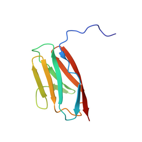
UniProt & NIH Common Fund Data Resources
Find proteins for Q14896 (Homo sapiens)
Explore Q14896 
Go to UniProtKB:  Q14896
PHAROS:  Q14896
GTEx:  ENSG00000134571 
Entity Groups
Sequence Clusters30% Identity50% Identity70% Identity90% Identity95% Identity100% Identity
UniProt GroupQ14896
Sequence Annotations
Expand
Reference Sequence

Experimental Data & Validation

Experimental Data

  • Method: SOLUTION NMR
  • Conformers Calculated: 100 
  • Conformers Submitted: 20 
  • Selection Criteria: target function 

Structure Validation

View Full Validation Report


Entry History 

Deposition Data

Revision History  (Full details and data files)

  • Version 1.0: 2009-03-17
    Type: Initial release
  • Version 1.1: 2011-07-13
    Changes: Version format compliance
  • Version 1.2: 2020-02-19
    Changes: Data collection, Database references, Derived calculations, Other
  • Version 1.3: 2024-05-01
    Changes: Data collection, Database references