Download Mendeley
NMR and X-RAY structures of human E2-like ubiquitin-fold modifier conjugating enzyme 1 (UFC1) reveal structural and functional conservation in the metazoan UFM1-UBA5-UFC1 ubiquination pathway.
Liu, G., Forouhar, F., Eletsky, A., Atreya, H.S., Aramini, J.M., Xiao, R., Huang, Y.J., Abashidze, M., Seetharaman, J., Liu, J., Rost, B., Acton, T., Montelione, G.T., Hunt, J.F., Szyperski, T.(2009) J Struct Funct Genomics 10: 127-136
- PubMed: 19101823 Search on PubMedSearch on PubMed Central
- DOI: https://doi.org/10.1007/s10969-008-9054-7
- Primary Citation Related Structures:
2K07, 3EVX - PubMed Abstract:
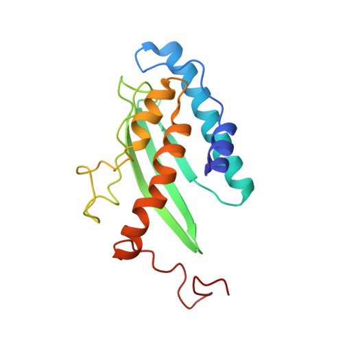
For cell regulation, E2-like ubiquitin-fold modifier conjugating enzyme 1 (Ufc1) is involved in the transfer of ubiquitin-fold modifier 1 (Ufm1), a ubiquitin like protein which is activated by E1-like enzyme Uba5, to various target proteins. Thereby, Ufc1 participates in the very recently discovered Ufm1-Uba5-Ufc1 ubiquination pathway which is found in metazoan organisms. The structure of human Ufc1 was solved by using both NMR spectroscopy and X-ray crystallography. The complementary insights obtained with the two techniques provided a unique basis for understanding the function of Ufc1 at atomic resolution. The Ufc1 structure consists of the catalytic core domain conserved in all E2-like enzymes and an additional N-terminal helix. The active site Cys(116), which forms a thio-ester bond with Ufm1, is located in a flexible loop that is highly solvent accessible. Based on the Ufc1 and Ufm1 NMR structures, a model could be derived for the Ufc1-Ufm1 complex in which the C-terminal Gly(83) of Ufm1 may well form the expected thio-ester with Cys(116), suggesting that Ufm1-Ufc1 functions as described for other E1-E2-E3 machineries. alpha-helix 1 of Ufc1 adopts different conformations in the crystal and in solution, suggesting that this helix plays a key role to mediate specificity.
- Department of Chemistry, Northeast Structural Genomics Consortium, The State University of New York at Buffalo, Buffalo, NY 14260, USA.
Organizational Affiliation: