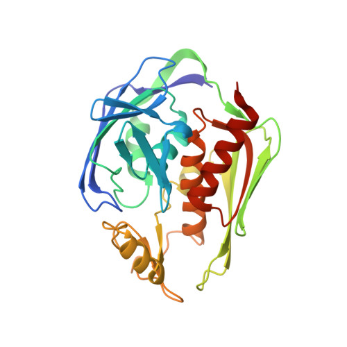
Structure of the deacetylase LpxC bound to the antibiotic CHIR-090: Time-dependent inhibition and specificity in ligand binding
Barb, A.W., Jiang, L., Raetz, C.R., Zhou, P.(2007) Proc Natl Acad Sci U S A 104: 18433-18438
- PubMed: 18025458
- DOI: https://doi.org/10.1073/pnas.0709412104
- Primary Citation of Related Structures:
2JT2 - PubMed Abstract:
The UDP-3-O-(R-3-hydroxyacyl)-N-acetylglucosamine deacetylase LpxC is an essential enzyme of lipid A biosynthesis in Gram-negative bacteria and a promising antibiotic target. CHIR-090, the most potent LpxC inhibitor discovered to date, displays two-step time-dependent inhibition and kills a wide range of Gram-negative pathogens as effectively as ciprofloxacin or tobramycin. In this study, we report the solution structure of the LpxC-CHIR-090 complex. CHIR-090 exploits conserved features of LpxC that are critical for catalysis, including the hydrophobic passage and essential active-site residues. CHIR-090 is adjacent to, but does not occupy, the UDP-binding pocket of LpxC, suggesting that a fragment-based approach may facilitate further optimization of LpxC inhibitors. Additionally, we identified key residues in the Insert II hydrophobic passage that modulate time-dependent inhibition and CHIR-090 resistance. CHIR-090 shares a similar, although previously unrecognized, chemical scaffold with other small-molecule antibiotics such as L-161,240 targeting LpxC, and provides a template for understanding the binding mode of these inhibitors. Consistent with this model, we provide evidence that L-161,240 also occupies the hydrophobic passage.
- Department of Biochemistry, Duke University Medical Center, Durham NC 27710.
Organizational Affiliation:


