2JJZ | pdb_00002jjz

Crystal Structure of Human Iba2, orthorhombic crystal form


Experimental Data Snapshot

  • Method: X-RAY DIFFRACTION
  • Resolution: 2.15 Å
  • R-Value Free: 
    0.257 (Depositor), 0.251 (DCC) 
  • R-Value Work: 
    0.216 (Depositor), 0.211 (DCC) 
  • R-Value Observed: 
    0.218 (Depositor) 

Starting Model: experimental
View more details

wwPDB Validation 3D Report Full Report

Validation slider image for 2JJZ

This is version 1.3 of the entry. See complete history

Literature

Structural and Functional Characterization of Human Iba Proteins.

Schulze, J.O.Quedenau, C.Roske, Y.Adam, T.Schuler, H.Behlke, J.Turnbull, A.P.Sievert, V.Scheich, C.Mueller, U.Heinemann, U.Bussow, K.

(2008) FEBS J 275: 4627

  • DOI: https://doi.org/10.1111/j.1742-4658.2008.06605.x
  • Primary Citation Related Structures: 
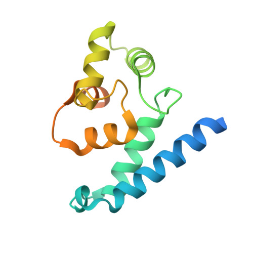
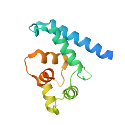
    2JJZ, 2VTG

  • PubMed Abstract: 

    Iba2 is a homolog of ionized calcium-binding adapter molecule 1 (Iba1), a 17-kDa protein that binds and cross-links filamentous actin (F-actin) and localizes to membrane ruffles and phagocytic cups. Here, we present the crystal structure of human Iba2 and its homodimerization properties, F-actin cross-linking activity, cellular localization and recruitment upon bacterial invasion in comparison with Iba1. The Iba2 structure comprises two central EF-hand motifs lacking bound Ca2+. Iba2 crystallized as a homodimer stabilized by a disulfide bridge and zinc ions. Analytical ultracentrifugation revealed a different mode of dimerization under reducing conditions that was independent of Ca2+. Furthermore, no binding of Ca2+ up to 0.1 mM was detected by equilibrium dialysis. Correspondingly, Iba EF-hand motifs lack residues essential for strong Ca2+ coordination. Sedimentation experiments and microscopy detected pronounced, indistinguishable F-actin binding and cross-linking activity of Iba1 and Iba2 with induction of F-actin bundles. Fluorescent Iba fusion proteins were expressed in HeLa cells and co-localized with F-actin. Iba1 was recruited into cellular projections to a larger extent than Iba2. Additionally, we studied Iba recruitment in a Shigella invasion model that induces cytoskeletal rearrangements. Both proteins were recruited into the bacterial invasion zone and Iba1 was again concentrated slightly higher in the cellular extensions.


  • Organizational Affiliation
    • Max Delbrück Center for Molecular Medicine, Berlin, Germany.

Macromolecule Content 

  • Total Structure Weight: 68.84 kDa 
  • Atom Count: 3,643 
  • Modeled Residue Count: 418 
  • Deposited Residue Count: 600 
  • Unique protein chains: 2

Macromolecules

Find similar proteins by:|  3D Structure
Entity ID: 1
MoleculeChains  Sequence LengthOrganismDetailsImage
IONIZED CALCIUM-BINDING ADAPTER MOLECULE 2150Homo sapiensMutation(s): 0 
UniProt & NIH Common Fund Data Resources
Find proteins for Q9BQI0 (Homo sapiens)
Explore Q9BQI0 
Go to UniProtKB:  Q9BQI0
PHAROS:  Q9BQI0
GTEx:  ENSG00000126878 
Entity Groups
Sequence Clusters30% Identity50% Identity70% Identity90% Identity95% Identity100% Identity
UniProt GroupQ9BQI0
Sequence Annotations
Expand
Reference Sequence
Find similar proteins by:|  3D Structure
Entity ID: 2
MoleculeChains  Sequence LengthOrganismDetailsImage
IONIZED CALCIUM-BINDING ADAPTER MOLECULE 2
B, C, D
150Homo sapiensMutation(s): 0 
UniProt & NIH Common Fund Data Resources
Find proteins for Q9BQI0 (Homo sapiens)
Explore Q9BQI0 
Go to UniProtKB:  Q9BQI0
PHAROS:  Q9BQI0
GTEx:  ENSG00000126878 
Entity Groups
Sequence Clusters30% Identity50% Identity70% Identity90% Identity95% Identity100% Identity
UniProt GroupQ9BQI0
Sequence Annotations
Expand
Reference Sequence

Small Molecules

Ligands 4 Unique
IDChains Name / Formula / InChI Key2D Diagram3D Interactions
ZN

Query on ZN


Download:Ideal Coordinates CCD File
E [auth A],
G [auth B],
I [auth C],
M [auth D]
ZINC ION
Zn
PTFCDOFLOPIGGS-UHFFFAOYSA-N
ACT

Query on ACT


Download:Ideal Coordinates CCD File
F [auth A],
J [auth C]
ACETATE ION
C2 H3 O2
QTBSBXVTEAMEQO-UHFFFAOYSA-M
CL

Query on CL


Download:Ideal Coordinates CCD File
H [auth B],
L [auth C]
CHLORIDE ION
Cl
VEXZGXHMUGYJMC-UHFFFAOYSA-M
NA

Query on NA


Download:Ideal Coordinates CCD File
K [auth C]SODIUM ION
Na
FKNQFGJONOIPTF-UHFFFAOYSA-N

Experimental Data & Validation

Experimental Data

  • Method: X-RAY DIFFRACTION
  • Resolution: 2.15 Å
  • R-Value Free:  0.257 (Depositor), 0.251 (DCC) 
  • R-Value Work:  0.216 (Depositor), 0.211 (DCC) 
  • R-Value Observed: 0.218 (Depositor) 
Space Group: P 21 21 2
Unit Cell:
Length ( Å )Angle ( ˚ )
a = 71.54α = 90
b = 186.5β = 90
c = 51.42γ = 90
Software Package:
Software NamePurpose
REFMACrefinement
XDSdata reduction
XSCALEdata scaling
PHASERphasing

Structure Validation

View Full Validation Report


Entry History 

Deposition Data

Revision History  (Full details and data files)

  • Version 1.0: 2009-07-14
    Type: Initial release
  • Version 1.1: 2011-07-13
    Changes: Advisory, Refinement description, Version format compliance
  • Version 1.2: 2023-12-13
    Changes: Data collection, Database references, Derived calculations, Other, Refinement description
  • Version 1.3: 2024-11-06
    Changes: Structure summary