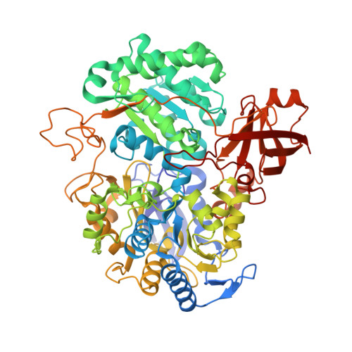
Periplasmic Nitrate Reductase Revisited: A Sulfur Atom Completes the Sixth Coordination of the Catalytic Molybdenum.
Najmudin, S., Gonzalez, P.J., Trincao, J., Coelho, C., Mukhopadhyay, A., Cerqueira, N.M.F.S.A., Romao, C.C., Moura, I., Moura, J.J.G., Brondino, C.D., Romao, M.J.(2008) J Biol Inorg Chem 13: 737
- PubMed: 18327621
- DOI: https://doi.org/10.1007/s00775-008-0359-6
- Primary Citation of Related Structures:
2JIM, 2JIO, 2JIP, 2JIQ, 2JIR, 2V3V, 2V45 - PubMed Abstract:
Nitrate reductase from Desulfovibrio desulfuricans ATCC 27774 (DdNapA) is a monomeric protein of 80 kDa harboring a bis(molybdopterin guanine dinucleotide) active site and a [4Fe-4S] cluster. Previous electron paramagnetic resonance (EPR) studies in both catalytic and inhibiting conditions showed that the molybdenum center has high coordination flexibility when reacted with reducing agents, substrates or inhibitors. As-prepared DdNapA samples, as well as those reacted with substrates and inhibitors, were crystallized and the corresponding structures were solved at resolutions ranging from 1.99 to 2.45 A. The good quality of the diffraction data allowed us to perform a detailed structural study of the active site and, on that basis, the sixth molybdenum ligand, originally proposed to be an OH/OH(2) ligand, was assigned as a sulfur atom after refinement and analysis of the B factors of all the structures. This unexpected result was confirmed by a single-wavelength anomalous diffraction experiment below the iron edge (lambda = 1.77 A) of the as-purified enzyme. Furthermore, for six of the seven datasets, the S-S distance between the sulfur ligand and the Sgamma atom of the molybdenum ligand Cys(A140) was substantially shorter than the van der Waals contact distance and varies between 2.2 and 2.85 A, indicating a partial disulfide bond. Preliminary EPR studies under catalytic conditions showed an EPR signal designated as a turnover signal (g values 1.999, 1.990, 1.982) showing hyperfine structure originating from a nucleus of unknown nature. Spectropotentiometric studies show that reduced methyl viologen, the electron donor used in the catalytic reaction, does not interact directly with the redox cofactors. The turnover signal can be obtained only in the presence of the reaction substrates. With use of the optimized conditions determined by spectropotentiometric titration, the turnover signal was developed with (15)N-labeled nitrate and in D(2)O-exchanged DdNapA samples. These studies indicate that this signal is not associated with a Mo(V)-nitrate adduct and that the hyperfine structure originates from two equivalent solvent-exchangeable protons. The new coordination sphere of molybdenum proposed on the basis of our studies led us to revise the currently accepted reaction mechanism for periplasmic nitrate reductases. Proposals for a new mechanism are discussed taking into account a molybdenum and ligand-based redox chemistry, rather than the currently accepted redox chemistry based solely on the molybdenum atom.
- Departamento de Química, FCT-UNL, REQUIMTE/CQFB, Monte de Caparica, 2829-516, Almada, Portugal. shabir@dq.fct.unl.pt
Organizational Affiliation:


