X-ray structure analysis of a metalloprotein with enhanced active-site resolution using in situ x-ray absorption near edge structure spectroscopy.
Arcovito, A., Benfatto, M., Cianci, M., Hasnain, S.S., Nienhaus, K., Nienhaus, G.U., Savino, C., Strange, R.W., Vallone, B., Della Longa, S.(2007) Proc Natl Acad Sci U S A 104: 6211-6216
- PubMed: 17404234 Search on PubMedSearch on PubMed Central
- DOI: https://doi.org/10.1073/pnas.0608411104
- Primary Citation Related Structures:
2JHO - PubMed Abstract:
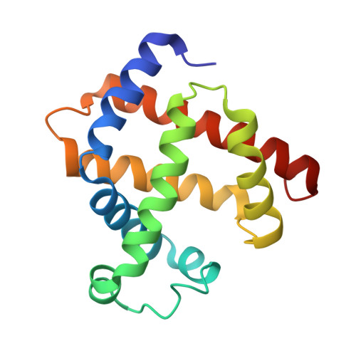
X-ray absorption spectroscopy is exquisitely sensitive to the coordination geometry of an absorbing atom and therefore allows bond distances and angles of the surrounding atomic cluster to be measured with atomic resolution. By contrast, the accuracy and resolution of metalloprotein active sites obtainable from x-ray crystallography are often insufficient to analyze the electronic properties of the metals that are essential for their biological functions. Here, we demonstrate that the combination of both methods on the same metalloprotein single crystal yields a structural model of the protein with exceptional active-site resolution. To this end, we have collected an x-ray diffraction data set to 1.4-A resolution and Fe K-edge polarized x-ray absorption near edge structure (XANES) spectra on the same cyanomet sperm whale myoglobin crystal. The XANES spectra were quantitatively analyzed by using a method based on the multiple scattering approach, which yielded Fe-heme structural parameters with +/-(0.02-0.07)-A accuracy on the atomic distances and +/-7 degrees on the Fe-CN angle. These XANES-derived parameters were subsequently used as restraints in the crystal structure refinement. By combining XANES and x-ray diffraction, we have obtained an cyanomet sperm whale myoglobin structural model with a higher precision of the bond lengths and angles at the active site than would have been possible with crystallographic analysis alone.
- Department of Biochemical Sciences and Consiglio Nazionale delle Ricerche, Institute of Molecular Biology and Pathology, University of Rome La Sapienza, P.le A. Moro 5, 00185 Rome, Italy.
Organizational Affiliation: