2JGS | pdb_00002jgs

Circular permutant of avidin


Experimental Data Snapshot

  • Method: X-RAY DIFFRACTION
  • Resolution: 1.90 Å
  • R-Value Free: 
    0.251 (Depositor), 0.247 (DCC) 
  • R-Value Work: 
    0.203 (Depositor), 0.202 (DCC) 
  • R-Value Observed: 
    0.206 (Depositor) 

Starting Model: experimental
View more details

wwPDB Validation 3D Report Full Report

Validation slider image for 2JGS

Ligand Structure Quality Assessment 


This is version 1.3 of the entry. See complete history

Literature

Rational Modification of Ligand-Binding Preference of Avidin by Circular Permutation and Mutagenesis.

Maatta, J.A.E.Airenne, T.T.Nordlund, H.R.Janis, J.Paldanius, T.A.Vainiotalo, P.Johnson, M.S.Kulomaa, M.S.Hytonen, V.P.

(2008) Chembiochem 9: 1124

  • DOI: https://doi.org/10.1002/cbic.200700671
  • Primary Citation Related Structures: 
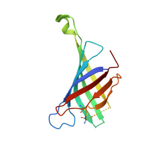
    2JGS

  • PubMed Abstract: 

    Chicken avidin is a key component used in a wide variety of biotechnological applications. Here we present a circularly permuted avidin (cpAvd4-->3) that lacks the loop between beta-strands 3 and 4. Importantly, the deletion of the loop has a positive effect on the binding of 4'-hydroxyazobenzene-2-carboxylic acid (HABA) to avidin. To increase the HABA affinity of cpAvd4-->3 even further, we mutated asparagine 118 on the bottom of the ligand-binding pocket to methionine, which simultaneously caused a significant drop in biotin-binding affinity. The X-ray structure of cpAvd4--> 3(N118M) allows an understanding of the effect of mutation to biotin-binding, whereas isothermal titration calorimetry revealed that the relative binding affinity of biotin and HABA had changed by over one billion-fold between wild-type avidin and cpAvd4-->3(N118M). To demonstrate the versatility of the cpAvd4-->3 construct, we have shown that it is possible to link cpAvd4-->3 and cpAvd5-->4 to form the dual-chain avidin called dcAvd2. These novel avidins might serve as a basis for the further development of self-organising nanoscale avidin building blocks.


  • Organizational Affiliation
    • Institute of Medical Technology, University of Tampere, 33014 University of Tampere, Finland.

Macromolecule Content 

  • Total Structure Weight: 58.21 kDa 
  • Atom Count: 3,792 
  • Modeled Residue Count: 450 
  • Deposited Residue Count: 520 
  • Unique protein chains: 1

Macromolecules

Find similar proteins by:|  3D Structure
Entity ID: 1
MoleculeChains  Sequence LengthOrganismDetailsImage
CIRCULAR PERMUTANT OF AVIDIN
A, B, C, D
130Gallus gallusMutation(s): 0 
UniProt
Find proteins for P02701 (Gallus gallus)
Explore P02701 
Go to UniProtKB:  P02701
Entity Groups
Sequence Clusters30% Identity50% Identity70% Identity90% Identity95% Identity100% Identity
UniProt GroupP02701
Sequence Annotations
Expand
Reference Sequence

Experimental Data & Validation

Experimental Data

  • Method: X-RAY DIFFRACTION
  • Resolution: 1.90 Å
  • R-Value Free:  0.251 (Depositor), 0.247 (DCC) 
  • R-Value Work:  0.203 (Depositor), 0.202 (DCC) 
  • R-Value Observed: 0.206 (Depositor) 
Space Group: P 1 21 1
Unit Cell:
Length ( Å )Angle ( ˚ )
a = 41.768α = 90
b = 79.309β = 98.73
c = 71.727γ = 90
Software Package:
Software NamePurpose
REFMACrefinement
XDSdata reduction
XDSdata scaling
PHASERphasing

Structure Validation

View Full Validation Report


Ligand Structure Quality Assessment 


Entry History 

Deposition Data

Revision History  (Full details and data files)

  • Version 1.0: 2008-03-04
    Type: Initial release
  • Version 1.1: 2011-07-13
    Changes: Refinement description, Version format compliance
  • Version 1.2: 2023-12-13
    Changes: Data collection, Database references, Other, Refinement description
  • Version 1.3: 2024-11-20
    Changes: Structure summary