Molecular Discrimination of Structurally Equivalent Lys 63-Linked and Linear Polyubiquitin Chains.
Komander, D., Reyes-Turcu, F., Licchesi, J.D.F., Odenwaelder, P., Wilkinson, K.D., Barford, D.(2009) EMBO Rep 10: 466
- PubMed: 19373254 Search on PubMedSearch on PubMed Central
- DOI: https://doi.org/10.1038/embor.2009.55
- Primary Citation Related Structures:
2JF5, 2W9N - PubMed Abstract:
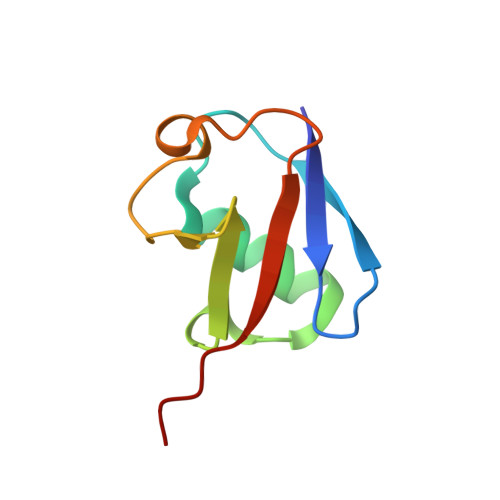
At least eight types of ubiquitin chain exist, and individual linkages affect distinct cellular processes. The only distinguishing feature of differently linked ubiquitin chains is their structure, as polymers of the same unit are chemically identical. Here, we have crystallized Lys 63-linked and linear ubiquitin dimers, revealing that both adopt equivalent open conformations, forming no contacts between ubiquitin molecules and thereby differing significantly from Lys 48-linked ubiquitin chains. We also examined the specificity of various deubiquitinases (DUBs) and ubiquitin-binding domains (UBDs). All analysed DUBs, except CYLD, cleave linear chains less efficiently compared with other chain types, or not at all. Likewise, UBDs can show chain specificity, and are able to select distinct linkages from a ubiquitin chain mixture. We found that the UBAN (ubiquitin binding in ABIN and NEMO) motif of NEMO (NF-kappaB essential modifier) binds to linear chains exclusively, whereas the NZF (Npl4 zinc finger) domain of TAB2 (TAK1 binding protein 2) is Lys 63 specific. Our results highlight remarkable specificity determinants within the ubiquitin system.
- Division of Protein and Nucleic Acid Chemistry, Medical Research Council Laboratory of Molecular Biology, Hills Road, Cambridge CB2 0QH, UK. dk@mrc-lmb.cam.ac.uk
Organizational Affiliation: