2J74 | pdb_00002j74

Structure of Beta-1,4-Galactanase


Experimental Data Snapshot

  • Method: X-RAY DIFFRACTION
  • Resolution: 2.60 Å
  • R-Value Free: 
    0.239 (Depositor) 
  • R-Value Work: 
    0.203 (Depositor) 
  • R-Value Observed: 
    0.203 (Depositor) 

Starting Model: experimental
View more details

wwPDB Validation 3D Report Full Report

Validation slider image for 2J74

This is version 2.1 of the entry. See complete history

Literature

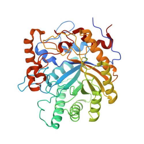
Investigating the Binding of Beta-1,4-Galactan to Bacillus Licheniformis Beta-1,4-Galactanase by Crystallography and Computational Modeling.

Le Nours, J.De Maria, L.Welner, D.Jorgensen, C.T.Christensen, L.L.H.Borchert, T.V.Larsen, S.Lo Leggio, L.

(2009) Proteins 75: 977

  • DOI: https://doi.org/10.1002/prot.22310
  • Primary Citation Related Structures: 
    2CCR, 2J74

  • PubMed Abstract: 

    Microbial beta-1,4-galactanases are glycoside hydrolases belonging to family 53, which degrade galactan and arabinogalactan side chains in the hairy regions of pectin, a major plant cell wall component. They belong to the larger clan GH-A of glycoside hydrolases, which cover many different poly- and oligosaccharidase specificities. Crystallographic complexes of Bacillus licheniformis beta-1,4-galactanase and its inactive nucleophile mutant have been obtained with methyl-beta(1-->4)-galactotetraoside, providing, for the first time, information on substrate binding to the aglycone side of the beta-1,4-galactanase substrate binding groove. Using the experimentally determined subsites as a starting point, a beta(1-->4)-galactononaose was built into the structure and subjected to molecular dynamics simulations giving further insight into the residues involved in the binding of the polysaccharide from subsite -4 to +5. In particular, this analysis newly identified a conserved beta-turn, which contributes to subsites -2 to +3. This beta-turn is unique to family 53 beta-1,4-galactanases among all clan GH-A families that have been structurally characterized and thus might be a structural signature for endo-beta-1,4-galactanase specificity.


  • Organizational Affiliation
    • Biophysical Chemistry Group, Department of Chemistry, University of Copenhagen, DK-2100 Copenhagen, Denmark.

Macromolecule Content 

  • Total Structure Weight: 89.33 kDa 
  • Atom Count: 6,214 
  • Modeled Residue Count: 775 
  • Deposited Residue Count: 798 
  • Unique protein chains: 1

Macromolecules

Find similar proteins by:|  3D Structure
Entity ID: 1
MoleculeChains  Sequence LengthOrganismDetailsImage
YVFO
A, B
399Bacillus licheniformisMutation(s): 0 
EC: 3.2.1.89 (PDB Primary Data), 3.2.1 (UniProt)
UniProt
Find proteins for Q65CX5 (Bacillus licheniformis (strain ATCC 14580 / DSM 13 / JCM 2505 / CCUG 7422 / NBRC 12200 / NCIMB 9375 / NCTC 10341 / NRRL NRS-1264 / Gibson 46))
Explore Q65CX5 
Go to UniProtKB:  Q65CX5
Entity Groups
Sequence Clusters30% Identity50% Identity70% Identity90% Identity95% Identity100% Identity
UniProt GroupQ65CX5
Sequence Annotations
Expand
Reference Sequence

Oligosaccharides

Help  
Entity ID: 2
MoleculeChains Length2D Diagram GlycosylationD Interactions
beta-D-galactopyranose-(1-4)-beta-D-galactopyranose-(1-4)-beta-D-galactopyranose
C, E
3N/A
Glycosylation Resources
GlyTouCan: G06996VN
GlyCosmos: G06996VN
GlyGen: G06996VN
Entity ID: 3
MoleculeChains Length2D Diagram GlycosylationD Interactions
beta-D-galactopyranose-(1-4)-beta-D-galactopyranose
D, F
2N/A
Glycosylation Resources
GlyTouCan: G08806VI
GlyCosmos: G08806VI
GlyGen: G08806VI

Small Molecules

Biologically Interesting Molecules (External Reference) 

2 Unique

Experimental Data & Validation

Experimental Data

  • Method: X-RAY DIFFRACTION
  • Resolution: 2.60 Å
  • R-Value Free:  0.239 (Depositor) 
  • R-Value Work:  0.203 (Depositor) 
  • R-Value Observed: 0.203 (Depositor) 
Space Group: P 1 21 1
Unit Cell:
Length ( Å )Angle ( ˚ )
a = 50.43α = 90
b = 80.25β = 99.5
c = 104.5γ = 90
Software Package:
Software NamePurpose
CNSrefinement
DENZOdata reduction
SCALEPACKdata scaling

Structure Validation

View Full Validation Report


Entry History 

Deposition Data

Revision History  (Full details and data files)

  • Version 1.0: 2007-12-18
    Type: Initial release
  • Version 1.1: 2011-05-08
    Changes: Version format compliance
  • Version 1.2: 2011-07-13
    Changes: Version format compliance
  • Version 1.3: 2018-01-17
    Changes: Data collection
  • Version 2.0: 2020-07-29
    Type: Remediation
    Reason: Carbohydrate remediation
    Changes: Atomic model, Data collection, Derived calculations, Non-polymer description, Structure summary
  • Version 2.1: 2023-12-13
    Changes: Data collection, Database references, Refinement description, Structure summary