2J6A | pdb_00002j6a

Structure of S. cerevisiae Trm112 protein, a methyltransferase activator


Experimental Data Snapshot

  • Method: X-RAY DIFFRACTION
  • Resolution: 1.70 Å
  • R-Value Free: 
    0.236 (Depositor), 0.236 (DCC) 
  • R-Value Work: 
    0.198 (Depositor), 0.197 (DCC) 
  • R-Value Observed: 
    0.200 (Depositor) 

wwPDB Validation 3D Report Full Report

Validation slider image for 2J6A

This is version 1.4 of the entry. See complete history

Literature

The Zinc Finger Protein Ynr046W is Plurifunctional and a Component of the Erf1 Methyltransferase in Yeast.

Heurgue-Hamard, V.Graille, M.Scrima, N.Ulryck, N.Champ, S.Van Tilbeurgh, H.Buckingham, R.H.

(2006) J Biological Chem 281: 36140

  • DOI: https://doi.org/10.1074/jbc.M608571200
  • Primary Citation Related Structures: 
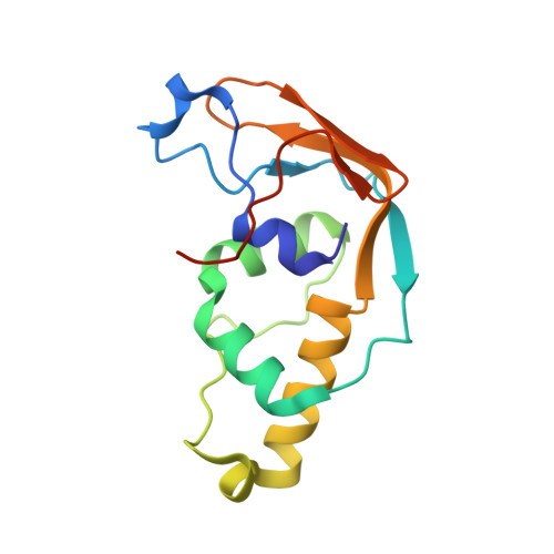
    2J6A

  • PubMed Abstract: 

    Protein release factor eRF1 in Saccharomyces cerevisiae, in complex with eRF3 and GTP, is methylated on a functionally crucial Gln residue by the S-adenosylmethionine-dependent methyltransferase Ydr140w. Here we show that eRF1 methylation, in addition to these previously characterized components, requires a 15-kDa zinc-binding protein, Ynr046w. Co-expression in Escherichia coli of Ynr046w and Ydr140w allows the latter to be recovered in soluble form rather than as inclusion bodies, and the two proteins co-purify on nickel-nitrilotriacetic acid chromatography when Ydr140w alone carries a His tag. The crystal structure of Ynr046w has been determined to 1.7 A resolution. It comprises a zinc-binding domain built from both the N- and C-terminal sequences and an inserted domain, absent from bacterial and archaeal orthologs of the protein, composed of three alpha-helices. The active methyltransferase is the heterodimer Ydr140w.Ynr046w, but when alone, both in solution and in crystals, Ynr046w appears to be a homodimer. The Ynr046w eRF1 methyltransferase subunit is shared by the tRNA methyltransferase Trm11p and probably by two other enzymes containing a Rossman fold.


  • Organizational Affiliation
    • UPR 9073 du CNRS, Institut de Biologie Physico-Chimique, 13 rue Pierre et Marie Curie, 75005 Paris, France.

Macromolecule Content 

  • Total Structure Weight: 16.03 kDa 
  • Atom Count: 1,215 
  • Modeled Residue Count: 136 
  • Deposited Residue Count: 141 
  • Unique protein chains: 1

Macromolecules

Find similar proteins by:|  3D Structure
Entity ID: 1
MoleculeChains  Sequence LengthOrganismDetailsImage
PROTEIN TRM112141Saccharomyces cerevisiaeMutation(s): 0 
EC: 2.1.1
UniProt
Find proteins for P53738 (Saccharomyces cerevisiae (strain ATCC 204508 / S288c))
Explore P53738 
Go to UniProtKB:  P53738
Entity Groups
Sequence Clusters30% Identity50% Identity70% Identity90% Identity95% Identity100% Identity
UniProt GroupP53738
Sequence Annotations
Expand
Reference Sequence

Experimental Data & Validation

Experimental Data

  • Method: X-RAY DIFFRACTION
  • Resolution: 1.70 Å
  • R-Value Free:  0.236 (Depositor), 0.236 (DCC) 
  • R-Value Work:  0.198 (Depositor), 0.197 (DCC) 
  • R-Value Observed: 0.200 (Depositor) 
Space Group: C 1 2 1
Unit Cell:
Length ( Å )Angle ( ˚ )
a = 92.6α = 90
b = 38.46β = 102.24
c = 45.81γ = 90
Software Package:
Software NamePurpose
REFMACrefinement
XDSdata reduction
XSCALEdata scaling
SHARPphasing

Structure Validation

View Full Validation Report


Entry History 

Deposition Data

Revision History  (Full details and data files)

  • Version 1.0: 2006-09-28
    Type: Initial release
  • Version 1.1: 2011-05-08
    Changes: Version format compliance
  • Version 1.2: 2011-07-13
    Changes: Version format compliance
  • Version 1.3: 2021-03-17
    Changes: Derived calculations, Other, Structure summary
  • Version 1.4: 2024-05-08
    Changes: Data collection, Database references