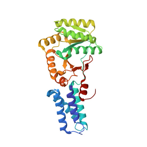
2J46

Water structure of T. Aquaticus Ffh NG Domain At 1.1A Resolution


Experimental Data Snapshot

  • Method: X-RAY DIFFRACTION
  • Resolution: 1.14 Å
  • R-Value Free: 0.172 
  • R-Value Work: 0.149 
  • R-Value Observed: 0.150 

Starting Model: experimental
View more details

wwPDB Validation   3D Report Full Report


This is version 1.3 of the entry. See complete history


Literature

Analysis of Protein Hydration in Ultra-High Resolution Structures of the Srp Gtpase Ffh

Ramirez, U.D.Freymann, D.M.

(2006) Acta Crystallogr D Biol Crystallogr 62: 1520

  • DOI: https://doi.org/10.1107/S0907444906040807
  • Primary Citation of Related Structures:  
    2J45, 2J46

  • PubMed Abstract: 

    Two new structures of the SRP GTPase Ffh have been determined at 1.1 A resolution and provide the basis for comparative examination of the extensive water structure of the apo conformation of these GTPases. A set of well defined water-binding positions have been identified in the active site of the two-domain ;NG' GTPase, as well as at two functionally important interfaces. The water hydrogen-bonding network accommodates alternate conformations of the protein side chains by undergoing local rearrangements and, in one case, illustrates binding of a solute molecule within the active site by displacement of water molecules without further disruption of the water-interaction network. A subset of the water positions are well defined in several lower resolution structures, including those of different nucleotide-binding states; these appear to function in maintaining the protein structure. Consistent arrangements of surface water between three different ultrahigh-resolution structures provide a framework for beginning to understand how local water structure contributes to protein-ligand and protein-protein binding in the SRP GTPases.


  • Organizational Affiliation

    Department of Molecular Pharmacology and Biological Chemistry, Northwestern University, Chicago, IL 60611, USA.


Macromolecules
Find similar proteins by:  (by identity cutoff)  |  3D Structure
Entity ID: 1
MoleculeChains Sequence LengthOrganismDetailsImage
SIGNAL RECOGNITION PARTICLE PROTEIN
A, B
297Thermus aquaticusMutation(s): 0 
EC: 3.6.5.4
UniProt
Find proteins for O07347 (Thermus aquaticus)
Explore O07347 
Go to UniProtKB:  O07347
Entity Groups  
Sequence Clusters30% Identity50% Identity70% Identity90% Identity95% Identity100% Identity
UniProt GroupO07347
Sequence Annotations
Expand
  • Reference Sequence
Small Molecules
Ligands 4 Unique
IDChains Name / Formula / InChI Key2D Diagram3D Interactions
MRD
Query on MRD

Download Ideal Coordinates CCD File 
C [auth A],
J [auth B]
(4R)-2-METHYLPENTANE-2,4-DIOL
C6 H14 O2
SVTBMSDMJJWYQN-RXMQYKEDSA-N
ACT
Query on ACT

Download Ideal Coordinates CCD File 
E [auth A],
K [auth B]
ACETATE ION
C2 H3 O2
QTBSBXVTEAMEQO-UHFFFAOYSA-M
MN
Query on MN

Download Ideal Coordinates CCD File 
H [auth A],
L [auth B]
MANGANESE (II) ION
Mn
WAEMQWOKJMHJLA-UHFFFAOYSA-N
CL
Query on CL

Download Ideal Coordinates CCD File 
D [auth A],
F [auth A],
G [auth A],
I [auth B]
CHLORIDE ION
Cl
VEXZGXHMUGYJMC-UHFFFAOYSA-M
Experimental Data & Validation

Experimental Data

  • Method: X-RAY DIFFRACTION
  • Resolution: 1.14 Å
  • R-Value Free: 0.172 
  • R-Value Work: 0.149 
  • R-Value Observed: 0.150 
  • Space Group: C 1 2 1
Unit Cell:
Length ( Å )Angle ( ˚ )
a = 109.473α = 90
b = 54.496β = 97.72
c = 98.799γ = 90
Software Package:
Software NamePurpose
REFMACrefinement
DENZOdata reduction
SCALEPACKdata scaling

Structure Validation

View Full Validation Report


Entry History 

Deposition Data

Revision History  (Full details and data files)

  • Version 1.0: 2006-11-30
    Type: Initial release
  • Version 1.1: 2011-05-08
    Changes: Version format compliance
  • Version 1.2: 2011-07-13
    Changes: Version format compliance
  • Version 1.3: 2023-12-13
    Changes: Data collection, Database references, Derived calculations, Other, Refinement description